


ibidi Material Specifications:
Choosing the Right Coverslip and Surface
Polymer or glass coverslip bottom—all ibidi labware is designed and optimized for both cell culture and high-quality microscopy.
Whether you select a polymer coverslip or a glass coverslip, you can rely on excellent optical performance and compatibility with a wide range of microscopy techniques. The main differences lie in the coatings and surface treatments, which are tailored to optimize cell adhesion, growth, and imaging.
On this page, you’ll find a clear comparison of the specifications and unique advantages of each option—helping you choose the best material for your specific research needs.
Quick Comparison of ibidi Bottoms
Choosing between polymer and glass coverslip bottoms depends on the requirements of your experiment:
|
| Standard polystyrene plates | ||||
| Bottom thickness | 180 µm (+10/–5 µm) | 170 µm (+/–5 µm) | 170 µm (+20/–10 µm) | 1000 µm (+/–50 µm) | 500–1000 µm | |
| Bottom material | Polymer | D 263 M Schott high precision glass | D 263 M Schott high precision glass | Soda-lime glass | Polymer | |
| Applications | Inverted high-resolution brightfield and fluorescence microscopy, live cell imaging | Inverted high-resolution, TIRF, super-resolution, live cell imaging | Inverted high-resolution brightfield and fluorescence microscopy, live cell imaging | Upright microscopy | Low-resolution routine microscopy |


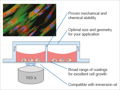
The ibidi µ-Slides, µ-Dishes, and µ-Plates are available with either a #1.5 ibidi Polymer Coverslip or a #1.5H Glass Coverslip bottom—both offering excellent optical clarity for standard microscopy techniques such as brightfield, phase contrast, and confocal microscopy. The tissue culture-treated ibiTreat Polymer Coverslip supports the growth of most cell types without any need for additional coatings and eases the use in cell microscopy.
While differences between the ibidi Polymer and Glass Coverslips are minimal in standard microscopy workflows, the choice of material can significantly affect advanced techniques such as TIRF and super-resolution microscopy.
For detailed optical specs, refer to our surface material guide below or evaluate the different coverslip materials through our Free Sample Program.
ibidi Surfaces and Coatings
The ibidi Polymer Coverslip provides optimal growth conditions for various cell-based assays and cell types and is available with different treatments or coatings (ibiTreat, Bioinert, Collagen IV, Poly-L-Lysine). You can find more information about the coatings in our Coating Guide. The glass coverslip is available as an untreated/uncoated version.

ibidi Polymer Coverslip
Available with different surfaces and coatings.
ibiTreat (Tissue Culture-Treated) Surface |
Hydrophobic, Untreated |
Bioinert Surface |
µ-Patterned Surface |
Coated Surface |

Glass Coverslip
Untreated and uncoated.
Glass Surface |
Microscopy Compatibility of Different Bottoms
Choosing the right coverslip bottom ensures optimal imaging quality for your experiment:
|
|
|
| Standard polystyrene plates & dishes*** | ||
| Brightfield Microscopy | ||||||
| Phase Contrast | ||||||
| Differential Interference Contrast (DIC) | ||||||
| Widefield Fluorescence | ||||||
| Confocal Fluorescence | ||||||
| Two-Photon and Mulitphoton Microscopy | ||||||
| Fluorescence Recovery After Photobleaching (FRAP) | ||||||
| Förster Resonance Energy Transfer (FRET) | ||||||
| Fluorescence Lifetime Imaging Microscopy (FLIM) | ||||||
| Lightsheet Fluorescence Microscopy (LSFM, SPIM) | ||||||
| Total Internal Reflection Fluorescence (TIRF) | ||||||
| Super-Resolution Microscopy |
= excellent / fully compatible, = usable with limitations, = not recommended
Optical Properties and Further Specifications of Different Bottoms
Optical specifications of the chosen material are crucial for the outcome of your experiment, especially for advanced microscopy applications:
|
|
|
| Standard polystyrene plates & dishes*** | ||
| Bottom thickness | 180 µm (+10/–5 µm) | 170 µm (+/–5 µm) | 170 µm (+20/–10 µm) | 1000 µm (+/–50 µm) | 500–1000 µm | |
| Bottom material | Polymer | D 263 M Schott high precision glass | D 263 M Schott high precision glass | Soda-lime glass | Polymer | |
| Refractive index (nD 589 nm) | 1.52 | 1.52 | 1.52 | 1.52 | 1.58 | |
| Abbe number | 56 | 55 | 55 | 64 | 31 | |
| Autofluorescence | No | No | No | No | Very high | |
| Transmission | Very high (even ultraviolet) | High (ultraviolet restrictions) | High (ultraviolet restrictions) | High (ultraviolet restrictions) | High (ultraviolet restrictions) | |
| Gas permeability | Yes | No | No | No | Yes | |
| Material flexibility | High | Low | Low | Very low | High | |
| Breakable | No | Yes | Yes | Yes | No | |
| Compatibility with protein coatings | Yes | Yes | Yes | Yes | Yes | |
| Birefringence (DIC) | Low (DIC-compatible) | Low (DIC-compatible) | Low (DIC-compatible) | Low (DIC-compatible) | Very high | |
| Immersion oil compatibility | Yes (compatibility list) | Yes | Yes | Yes | No | |
| Correct thickness for high-resolution microscopy objective lenses | Yes | Yes | Yes | No (Yes, if mounted with a standard glass coverslip) | No |
** The Glass Bottom Dish 35 mm is produced by using a standard #1.5 glass coverslip with a thickness of 170 µm (+20 µm/–10 µm). This glass bottom material fulfills the needs of all standard applications where a cost-effective coverslip is required.
*** With the 3 Well | 8 Well | 12 Well Chamber Slide, removable, ibidi provides self-adhesive, removable silicone chambers that are mounted on a standard glass slide. They are suitable for upright and inverted microscopy (after mounting with a coverslip) and enable long-term storage of samples after immunofluorescence staining.
*** Not offered by ibidi.