


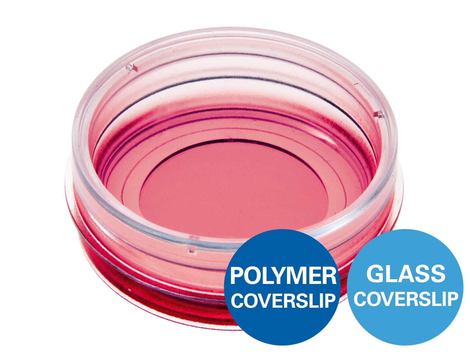
Suspension Cell Culture and Imaging
Several ibidi products are suitable for microscopy-based assays with suspension cells and other non-adherent cells. Depending on the application, suspension cells can either be confined in small wells or embedded in a 3D gel matrix, enabling controlled liquid handling, medium exchange, live cell imaging, and cell migration assays.
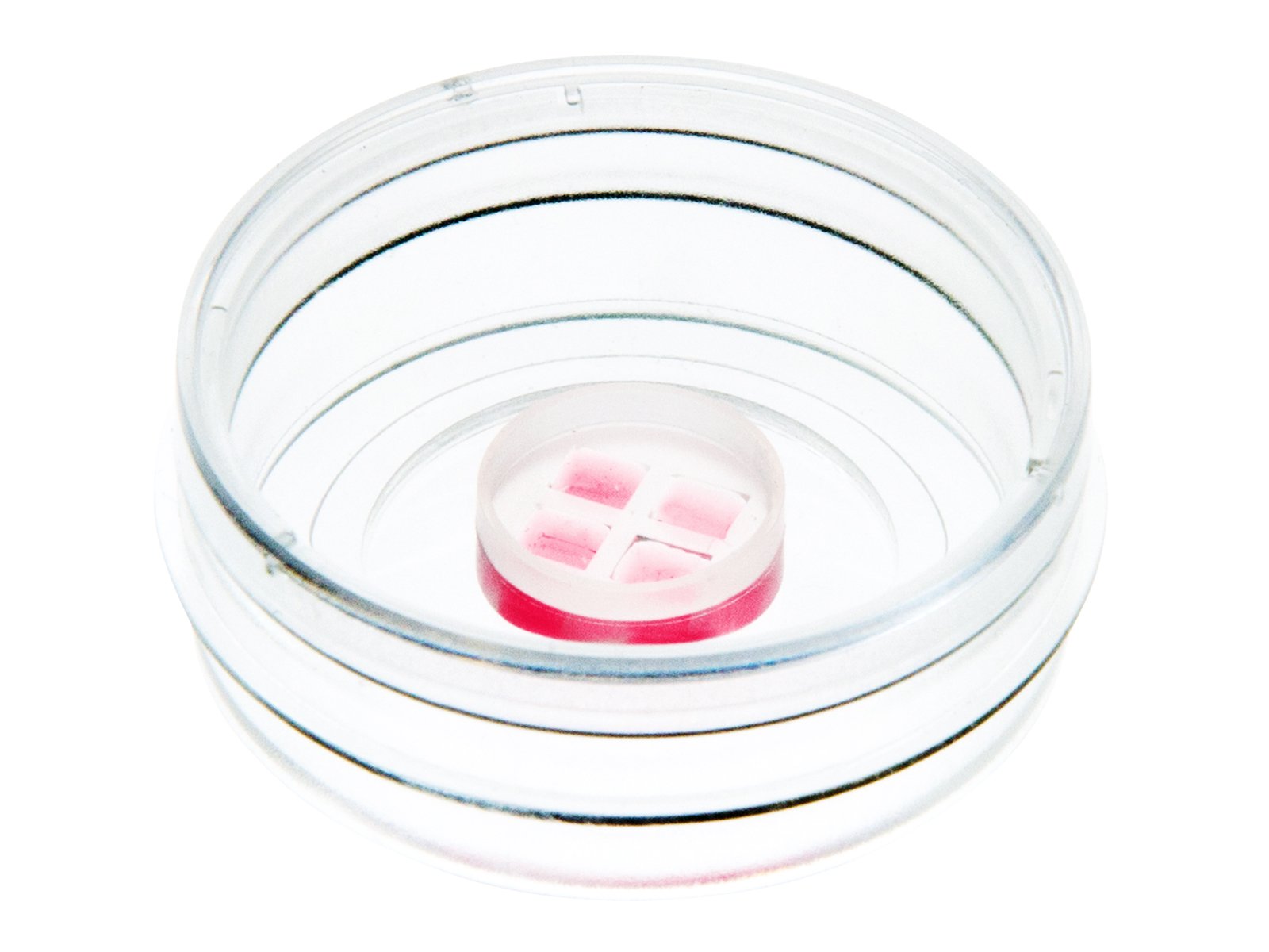
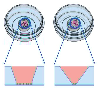
The micro-Insert 4 Well facilitates suspension cell handling without a gel matrix. The cells settle at the bottom of the four 10 µl conical wells, which keep small numbers of non-adherent cells within a defined observation area during microscopy. Medium can be carefully exchanged in the surrounding µ-Dish while the cells remain confined within the wells.
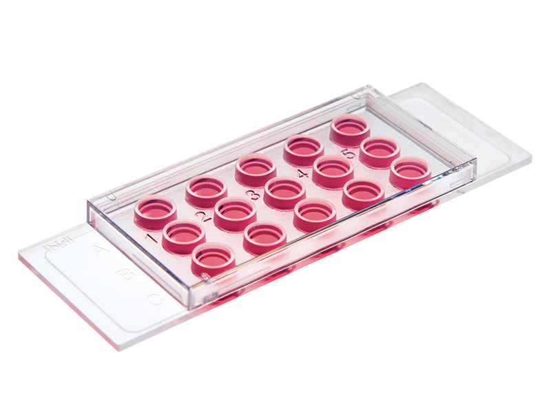
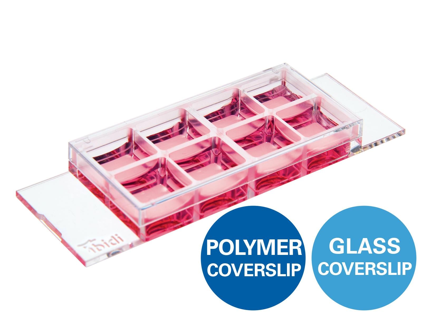
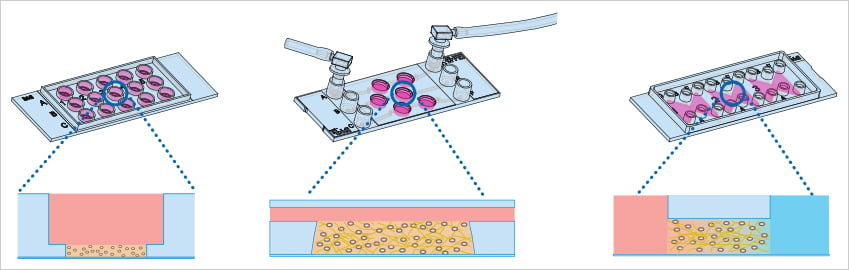
With the µ-Slide 15 Well 3D, suspension cells can be embedded in 10 µl of a 3D gel matrix. The cells can be cultured and imaged within the gel, while the overlying culture medium can be exchanged to support extended live cell imaging experiments.
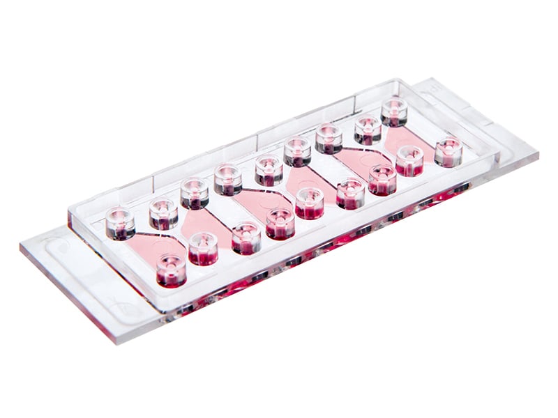
In the µ-Slide III 3D Perfusion, suspension cells can be embedded in a gel matrix within the 30 µl wells. The wells are connected to channels that enable gentle medium perfusion, allowing nutrients, compounds, or staining solutions to be delivered to the 3D cell culture.
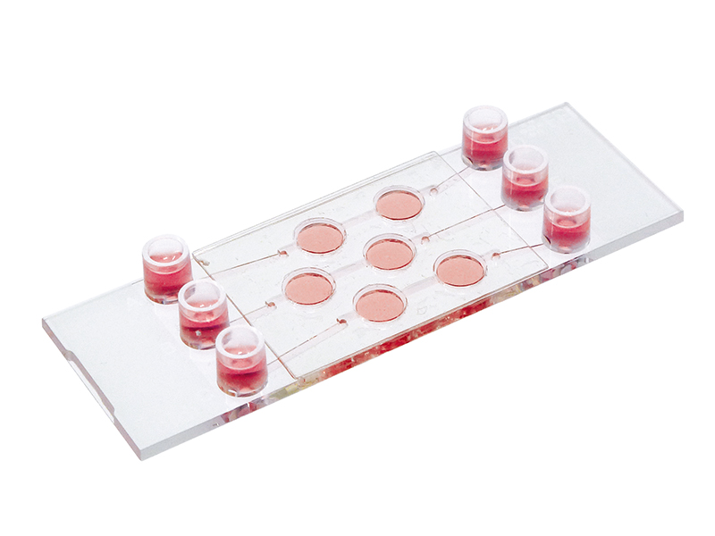
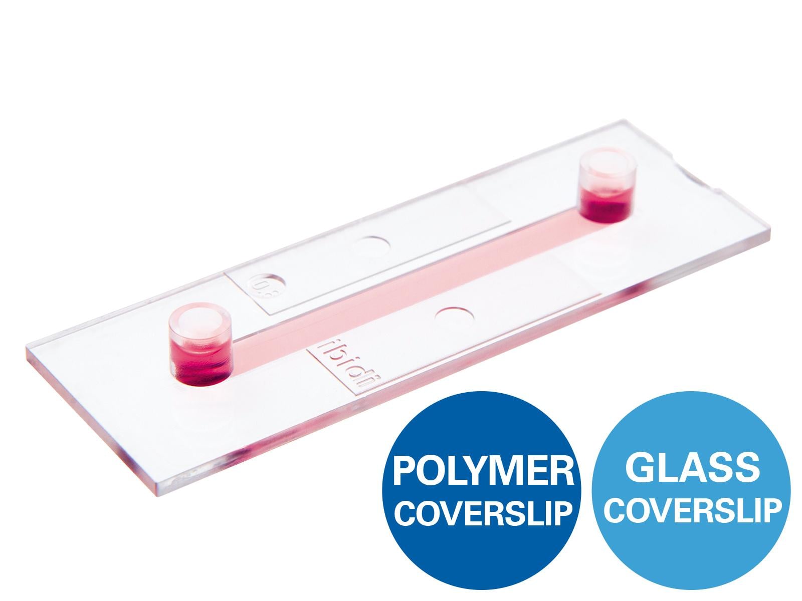
For studying suspension cell migration and chemotaxis in a 3D gel matrix, the µ-Slide Chemotaxis can be used. Suspension cells are mixed with a gel matrix and introduced into the observation area between two reservoirs. A chemoattractant added to one reservoir forms a stable chemical gradient by diffusion through the gel, enabling live cell imaging and analysis of directed cell migration.
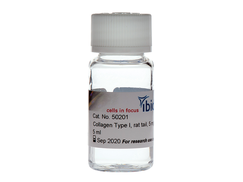
For embedding suspension cells in 3D gel matrices, we recommend Collagen Type I, Matrigel®, or another suitable hydrogel, depending on the cell type and application.