


Immune Cell Analysis
Innate and adaptive immune responses both depend on leukocytes, also known as white blood cells. They are produced in the bone marrow and circulate in the bloodstream, helping to defend the body against infection and disease. Leukocytes are involved in a wide range of immune processes, from the initial recognition of pathogens to the destruction and removal of infected cells.
There are several different types of leukocytes, each with its specific immune system function. Some of them, such as neutrophils and macrophages, help to engulf and destroy invading pathogens. Lymphocytes are a specialized subgroup of leukocytes that include B cells and T cells, which are crucial during the adaptive immune response. While B cells produce antibodies that can recognize and neutralize specific pathogens, T cells help to identify and destroy infected or abnormal cells in the body.
Multiple factors can affect leukocyte numbers and function, including age, stress, and disease. Live cell imaging of immune cells in a physiologic environment can give many insights into cell-cell interactions, cell death, proliferation, cytoskeleton dynamics, and many more aspects.
Immune cell markers are proteins on the surface of immune cells, which help to identify and distinguish different types of cells in the immune system. By analyzing the expression of these markers and further relevant proteins by immunofluorescence staining, researchers can help increase knowledge of the function and behavior of immune cells in health and disease.
These are only two methods that are instrumental in studying immune function and developing immunotherapies for a wide range of diseases, including cancer and autoimmune disorders.
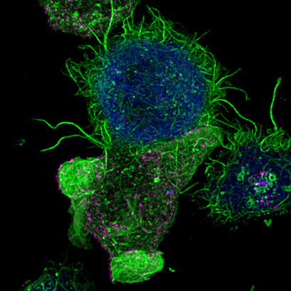
Confocal image of a Jurkat T lymphocyte (bottom) forming an immunological synapse with an antigen-presenting cell (top, blue). Filamentous actin is visualized in green. Multivesicular bodies in magenta. Cells were imaged using a µ-Slide 8 Well. Image by Manuel Izquierdo, IIBM, CSIC, Madrid, Spain.
Find Out MorePlease find more detailed information about the planning, conduction, and data analysis of immunofluorescence assays or live cell imaging experiments in our Application Chapters. |
|

ibidi Solutions for Immune Cell Analysis
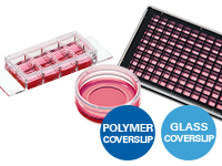
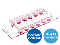
The ibidi µ-Slides and µ-Dishes include different geometries that combine optimal conditions for everyday cell culture and functional cell-based assays. They are ideal for immunofluorescence, live cell imaging, and high-resolution microscopy. The ibidi labware is available with the ibidi Polymer Coverslip and the ibidi Glass Coverslip. |
|
The ibidi channel slides, especially the µ-Slide VI 0.4, are particularly suitable for immunofluorescence stainings: Their geometry is ideal for the exact exchange of small medium amounts, which is necessary during immunocytochemistry stainings. The channel geometry is ideal for low-volume immunofluorescence assays. |
|
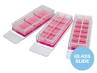
The Chamber Slides, removable are ideal for both low- and high-throughput immunofluorescence experiments and are suitable for the long-term storage of samples that are mounted with a glass coverslip. |
|
The ibidi µ-Plates are ideal for high throughput drug screening and large-scale knock-down and overexpression experiments with high-resolution microscopy as readout. These imaging plates are compatible with robotics and plate readers due to an ANSI/SLAS (SBS) standard format. The ibidi µ-Plates have 24, 96, and 384 wells and are available with the ibidi Polymer Coverslip and the ibidi Glass Coverslip with extremely low autofluorescence for undisturbed fluorescence microscopy. |
|
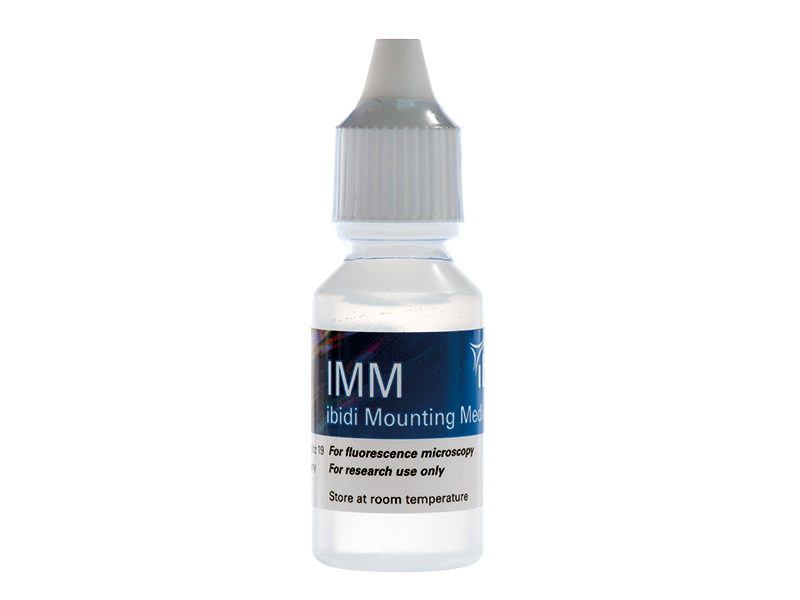
The ibidi Mounting Medium and the ibidi Mounting Medium With DAPI for immunofluorescence have a very low autofluorescence, prevent photobleaching and allow the sample to be stored for several weeks on the µ-Slide without the need for additional coverslips. |
|
The ibidi Stage Top Incubators provide physiological conditions for live cell imaging on every standard inverted microscope. They include CO2 and O2 control (e.g., for hypoxia experiments) as well as actively controlled humidity. They are available for single slides, dishes, and multiwell plates. |
|

Selected References
Live cell imaging for the measurement of cell death of T cells and monocyte-derived macrophages (MDMs) in response to different treatments using 4 Well FulTrac microinserts placed into a µ-Plate 24 Well Black
Linder A, Bauernfried S, Cheng Y, et al. CARD8 inflammasome activation triggers pyroptosis in human T cells. EMBO J. 2020;39(19):e105071. doi:10.15252/embj.2020105071.
Read article
Imaging of cytoskeleton dynamics of cytotoxic T lymphocytes (CTLs) in the µ-Slide 8 Well Glass Bottom using total internal reflection fluorescence (TIRF) microscopy and confocal laser scanning microscopy (CLSM)
Klein-Hessling S, Muhammad K, Klein M, et al. NFATc1 controls the cytotoxicity of CD8+ T cells. Nat Commun. 2017;8(1):511. Published 2017 Sep 11. doi:10.1038/s41467-017-00612-6.
Read article
Examination of neutrophil interactions with C. albicans biofilms using immunofluorescence microscopy in the µ-Slide 8 Well
Kernien JF, Johnson CJ, Bayless ML, Chovanec JF, Nett JE. Neutrophils From Patients With Invasive Candidiasis Are Inhibited by Candida albicans Biofilms. Front Immunol. 2020;11:587956. Published 2020 Dec 3. doi:10.3389/fimmu.2020.587956.
Read article
Read on and learn more about methods for analyzing the Tumor Microenvironment, Immune Cell Migration and Chemotaxis, or Immune Cell Cultivation Under Shear Stress.