Application Notes: Imaging and Microscopy
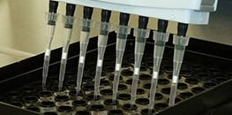
AN 05: Tube Formation Assay in the µ-Plate 96 Well 3D (PDF)
Handling protocol for tube formation assays using a multi-channel pipette and the µ-Plate 96 Well 3D
AN 12: Avoiding Evaporation: Humidity Control in Cell Culture (PDF)
An overview of different methods to avoid evaporation from cell culture vessels by controlling the humidity
AN 14: Live Cell Imaging Under Flow (PDF)
Discussion of various setups for live cell imaging experiments under flow conditions
AN 37: Image Shift Correction in Microscopic Time Lapse Sequences (PDF)
Instructions for correcting an externally generated shift of the sample in time lapse images (e.g. in chemotaxis experiments)
AN 02: Fluorescence Staining Using a µ-Slide I (PDF)
Examples describing how to do immunofluorescence stainings using µ-Slides
AN 09: Fluorescence Staining Using a µ-Slide VI (PDF)
Examples describing how to do immunofluorescence stainings using µ-Slides
AN 15: Fluorescence Staining Using the µ-Slide y-shaped (PDF)
Examples describing how to do immunofluorescence stainings using µ-Slides
AN 16: Immunofluorescence Staining Using the µ-Slide 8 Well high (PDF)
Detailed information on how to perform an immunofluorescence (IF) staining in the µ-Slide 8 Well high
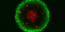
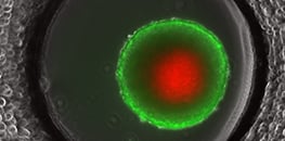
AN 33: Live / Dead Staining with FDA and PI (PDF)
Viability staining of adherent cells, single cells embedded in extracellular matrix, and cellular clusters
AN 44: Immunofluorescence Staining of HUVEC in 3D in the μ-Slide Chemotaxis (PDF)
A detailed protocol for immunostaining of HUVEC in a gel matrix in the µ-Slide Chemotaxis.
AN 45: Mounting Medium Types (PDF)
A comparison of non-hardening and hardening mounting media
AN 49: Fluorescence Staining Using a 12 Well Chamber, Removable (PDF)
A handling protocol for immunofluorescence staining in the 12 Well Chamber, removable
AN 50: Fluorescence Staining Using a 3 Well Chamber, Removable (PDF)
A handling protocol for immunofluorescence staining in the 3 Well Chamber, removable, including instructions for using the volume-minimizing coverslip
AN 58: Immunofluorescence Staining Using the μ-Slide 18 Well (PDF)
Example for an immunofluorescence staining in the µ-Slide 18 Well.
AN 61: Immunofluorescence Staining of HUVECs on a Gel Matrix Using the µ-Slide I Luer 3D (PDF)
Immunofluorescence staining of human endothelial cells on a gel matrix using the μ-Slide I Luer 3D
AN 64: FDA/PI Live/Dead Staining Using L929 Spheroids in the µ Slide Spheroid Perfusion (PDF)
Protocol for an FDA/PI fluorescence staining in the µ-Slide Spheroid Perfusion to distinguish living and dead cells in a spheroid
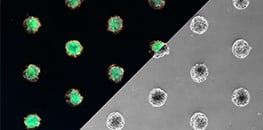
AN 78: Cell Culture and Immunofluorescence Staining in the μ-Slide VI 0.4 μ-Pattern ibiTreat (PDF)
A protocol for the cultivation, fixation, and staining of cells or spheroids on micropatterns
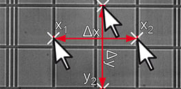
AN 22: Determination of the Pixel Size in Microscopy Images (PDF)
Measuring and calculating the pixel size of microscopic images
AN 37: Image Shift Correction in Microscopic Time Lapse Sequences (PDF)
Instructions for correcting an externally generated shift of the sample in time lapse images (e.g. in chemotaxis experiments)
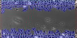
AN 67: Data Analysis of Wound Healing and Cell Migration Assays (PDF)
Methods for data analysis of wound healing assays and cell migration assays
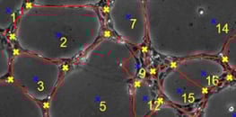
AN 70: Data Analysis of Tube Formation Assays (PDF)
Protocol for data analysis of tube formation assays


