Neurobiology

Molecular and cellular neurobiology research focuses on the development, organization, and function of the nervous system. Questions about how the neural stem cells develop into neurons or how the neurons generate and transmit signals, among other aspects, are addressed.

Since the early days of neurobiological research, microscopy has been a critical technique for gaining insight into the structural and functional properties of the nervous system. While the preparation and imaging of histological samples (e.g., brain slices) were already possible in the late 19th century, the development of advanced imaging techniques over the last 50 years, such as confocal microscopy and 2-photon microscopy, have advanced the visualization of neuronal structures and processes. The combination of new fluorescent probes and labeling techniques have made it possible to depict different neuronal components, and their complex interaction networks, in fixed and even living cells, tissues, or organisms.

Neurons differentiated from neural crest stem cells. Neurons are stained in green, nuclei in blue, and supporting glial cells in red.
Courtesy: Xue Shifeng, Institute of Medical Biology, A*STAR, Singapore


Most recently, with the invention of super-resolution microscopy and its ability to overcome the diffraction barrier of the light (~200nm), structures even as small as synaptic vesicles (with a size of 40nm) can be visualized.

In summary, the recent advancements in imaging techniques have allowed for the visualization of neuronal processes and morphology with high spatiotemporal resolution, which play a major role in understanding the function of the brain and nerves.

ibidi has contributed to these progressions by developing many specialized solutions for a variety of neurobiology-related, microscopy-based applications. More than 7000 published neuronal research papers mention ibidi applications in their references.

These articles cite products, such as labware with various geometries and coatings, as providing the ideal surface for cultivating and imaging even difficult to culture neuronal cells. Additionally, the ibidi Stage Top Incubator and Pump System were significant tools that allowed for the cultivation of cells under physiological conditions, thus making long-term, live-cell imaging of neuronal cells possible.

ibidi Products for Neurobiology Research

µ-Slide 8 Well | µ-Slide 8 Well high

  • A chambered coverslip with 8 wells for everyday cell culture, immunofluorescence, and high-end microscopy; suitable for a variety of neurobiology-related assays. Now, also available as the µ-Slide 8 Well high with individual wells and higher walls.

S_8082X_Slide_8well_8well_high_RGB_2Icon

Find more details and ordering information for the µ-Slide 8 Well and µ-Slide 8 Well high.


Rat fibroblast, surrounded by parallel-aligned rat Schwann cells (SCs), cultured in an ibidi µ-Slide 8 Well and stained for the SC-marker S100 (green), Vimentin (grey), and DAPI (blue). The image was obtained with a laser-scanning microscope.
Courtesy: Flavia Millesi, Medical University Vienna, Austria


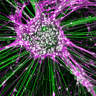
Rat dorsal root ganglionic cells and Schwann cells, cultured in an ibidi μ-Slide 8 Well, and stained for neurofilament (green), NGFR (magenta), and DAPI (white). The image was obtained with a LEICA SP8X laser scanning microscope.
Courtesy: Tamara Weiss, Division of Plastic and Reconstructive Surgery, Medical University of Vienna, Austria

Customer Comment

Federica Bono, University of Brescia, Italy

The µ-Slide 8 Well high worked very well, especially for our iPSC-derived neurons. We used them for live cell imaging microscopy, and we had very good results. We are very satisfied with the slide!

Federica Bono, University of Brescia, Italy

Selected References

Imaging of 3D Neuronal Co-Cultures

The ibidi µ-Slide 8 Well was used to image 3D cultures of neurons and meningeal fibroblasts in hydrogels.

T. E. Santos, B. Schaffran, N. Broguière, L. Meyn, M. Zenobi-Wong, F. Bradke. Axon Growth of CNS Neurons in Three Dimensions Is Amoeboid and Independent of Adhesions. Cell Reports, 2021, 10.1016/j.celrep.2020.107907.
Read article

Imaging of Humanized Three-Dimensional Peripheral Neurovascular (PNV) Models

The µ-Slide 8 Well was used to image a 3D co-culture model comprised of human embryonic stem cell peripheral sensory neurons (hESC)(PSNs) and hESC endothelial cells mixed with collagen type 1.

S. Kannan, M. Lee, S. Muthusamy, A. Blasiak, G. Sriram, T. Cao. Peripheral sensory neurons promote angiogenesis in neurovascular models derived from hESCs. Stem Cell Research, 2021, 10.1016/j.scr.2021.102231.
Read article

Live Cell Imaging of Cortical Neurons

The µ-Slide 8 Well was used for confocal live-cell imaging of Purkinje neurons.

E. Motori, I. Atanassov, S. M. V. Kochan, K. Folz-Donahue, V. Sakthivelu, P. Giavalisco, N. Toni, J. Puyal, N.-G. Larsson. Neuronal metabolic rewiring promotes resilience to neurodegeneration caused by mitochondrial dysfunction. Science Advances, 2020, 10.1126/sciadv.aba8271.
Read article

Immunofluorescence of Neuronal Cells

The µ-Slide 8 Well was used for the cultivation and immunostaining of rat Schwann cells (rSC), fibroblasts (rFB), and dorsal root ganglion (rDRG) neurons.

F . Millesi, T . Weiss, A. Mann, M. Haertinger, L. Semmler, P. Supper, D.Pils, A. Naghilou, C. Radtke. Defining the regenerative effects of native spider silk fibers on primary Schwann cells, sensory neurons, and nerve-associated fibroblasts. FASEB Journal. 2021, 10.1096/fj.202001447R
Read article

Immunofluorescence and Super-Resolution of Primary Hippocampal Neurons

The µ-Slide 8 Well was used for the immunofluorescence staining of primary hippocampal neurons, and subsequent confocal imaging and structure illumination microscopy (SIM).

L. Colnaghi, A. Conz, L. Russo, C.A. Musi, L. Fioriti, T. Borsello, M. Salmona. Neuronal Localization of SENP Proteins with Super Resolution Microscopy. Brain Sciences, 2020, 10.3390/brainsci10110778
Read article

µ-Slide 2 Well

  • A chambered coverslip with 2 wells, for use in immunofluorescence and high-end microscopy; suitable for a variety of neurobiology-related assays

Selected Reference

Growth Experiments and Live-Cell Imaging of Neuronal Cells
The µ-Slide 2 Well was used to compare different growth substrates for rat Schwann cells (rSC), fibroblasts (rFB), and dorsal root ganglion (rDRG) neurons. In addition, the µ-Slide 2 Well was used for the live-cell imaging of rSCs, rFBs, and rSCs.

F . Millesi, T . Weiss, A. Mann, M. Haertinger, L. Semmler, P. Supper, D.Pils, A. Naghilou, C. Radtke. Defining the regenerative effects of native spider silk fibers on primary Schwann cells, sensory neurons, and nerve-associated fibroblasts. FASEB J. 2021, 10.1096/fj.202001447R
Read article

Find more details and ordering information here.

µ-Slide 4 Well

  • A chambered coverslip with 4 wells, for use in immunofluorescence and high-end microscopy; suitable for a variety of neurobiology-related assays

Mouse primary skeletal muscle fibers (visualized by Desmin staining in green) in co-culture with primary motor neurons (visualized by SMI-32 staining in red), imaged in an ibidi µ-Slide 4 Well chambered coverslip. Nuclei are stained with DAPI (blue).

Courtesy: A. Ghasemizadeh, Institut NeuroMyoGène, Lyon University

Find more details and ordering information here.

µ-Slide I Luer

  • Channel slides with different heights, volumes, and coatings that are specially suited for flow applications; suitable for a variety of neurobiology-related assays

Selected Reference

Cultivation of Brain Microvascular Endothelial Cells (BMECs) Under Flow
To investigate the effect of shear stress on gerbil brain microvascular endothelial cells (BMECs), the cells were seeded into the µ-Slide I 0.6 Luer, then subjected to unidirectional flow with the ibidi Pump System.

J. Q.Gao, P. Wang, J. W. Yan, L. N. Ba, P. L. Shi, H. M. Wu, X. Y. Guan, Y. G. Cao, H.L. Sun, X. Y. Mao. Shear Stress Rescued the Neuronal Impairment Induced by Global Cerebral Ischemia Reperfusion via Activating PECAM-1-eNOS-NO Pathway. Frontiers in cell and developmental biology, 2021, 10.3389/fcell.2020.631286
Read article

Find more details and ordering information here.

µ-Dish 35 mm, high

  • A 35 mm imaging dish with a polymer coverslip bottom for high-end microscopy and cell-based assays; suitable for a variety of neurobiology-related assays

Selected References

Imaging of Neuronal CAD Cells
The µ-Dish 35 mm, high was used to culture and immunostain neuronal CAD (Cath.a-differentiated) cells, in order to quantify tunneling nanotubes connected cells (TNT), and filopodia connected cells, as well as to image the transfer of DiD-labelled vesicles between two cell populations.

B. Bhat, N. Ljubojevic, S. Zhu, M. Fukuda, A. Echard, C. Zurzolo. Rab35 and its effectors promote formation of tunneling nanotubes in neuronal cells. Scientific reports, 2020, 10.1038/s41598-020-74013-z
Read article

Calcium Imaging of Neuronal Cells
The µ-Dish 35 mm, high was used for Calcium imaging to evaluate cell functionality after differentiating human induced pluripotent stem cells (iPSCs) into neurons.

F. Bianchi, M. Malboubi, Y. Li, J. H. George, A. Jerusalem, F. Szele, M. S. Thompson, H. Ye. Rapid and efficient differentiation of functional motor neurons from human iPSC for neural injury modelling. Stem Cell Research, 2018, 10.1016/j.scr.2018.09.006.
Read article

Find more details and ordering information here.

µ-Slide Chemotaxis

  • A microslide developed to investigate chemotaxis of fast or slow migrating adherent cells and non-adherent cells in 2D or 3D gel matrices; also suitable for neuronal cell applications, such as monitoring axon outgrowth

Selected Reference

Monitoring Axon Outgrowth
The µ-Slide Chemotaxis was used to follow axon outgrowth in a 3D gel, in response to a chemical stimulus with live-cell microscopy.

D. Terheyden-Keighley, B. Brand-Saberi, C. Theiss. Real-Time Imaging of Accessible Axon Guidance Assays in Three-Dimensional Culture. Journal of Neurology and Experimental Neuroscience, 2017, 10.17756/jnen.2016-015
Read article

Find more details and ordering information here.

ibidi Pump System

  • A pump system for the cultivation of (neuronal) cells under flow; for simulation of blood vessels and various physiological conditions

Selected References

Cultivation of Brain Microvascular Endothelial Cells (BMECs) Under Flow
To investigate the effect of shear stress on gerbil brain microvascular endothelial cells (BMECs), cells were seeded into the µ-Slide I 0.6 Luer and subjected to unidirectional flow with the ibidi Pump System.

J. Q. Gao, P. Wang, J. W. Yan, L. N. Ba, P. L. Shi, H. M. Wu, X. Y. Guan, Y. G. Cao, H.L. Sun, X. Y. Mao. Shear Stress Rescued the Neuronal Impairment Induced by Global Cerebral Ischemia Reperfusion via Activating PECAM-1-eNOS-NO Pathway. Frontiers in cell and developmental biology, 2021, 10.3389/fcell.2020.631286
Read article

Cultivation of Astrocytes Under Flow
Astrocytes were incubated at physiological conditions using the ibidi Stage Top Incubator and exposed to shear stress using the ibidi Pump System.

N.M. Wakida, G.M.S. Cruz, P. Pouladian, M.W. Berns, D. Preece. Fluid Shear Stress Enhances the Phagocytic Response of Astrocytes. Front Bioeng Biotechnol, 2020, 10.3389/fbioe.2020.596577.
Read article

Find more details and ordering information here.

ibidi Stage Top Incubator

  • A Stage Top Incubator designed for easy live cell imaging on every inverted microscope; Universal Fit with CO2 control

Selected References

Live Cell Imaging of Neuronal Cells
DIC live cell imaging of central nervous system (CNS) neurons was performed on the ibidi Stage Top Incubator.

T. E. Santos, B. Schaffran, N. Broguière, L. Meyn, M. Zenobi-Wong, F. Bradke. Axon Growth of CNS Neurons in Three Dimensions Is Amoeboid and Independent of Adhesions. Cell Reports, 2021, 10.1016/j.celrep.2020.107907.
Read article

Cultivation of Astrocytes Under Physicological Conditions
Astrocytes were incubated at physiological conditions using the ibidi Stage Top Incubator and exposed to shear stress using the ibidi Pump System.

N.M. Wakida, G.M.S. Cruz, P. Pouladian, M.W. Berns, D. Preece. Fluid Shear Stress Enhances the Phagocytic Response of Astrocytes. Front Bioeng Biotechnol, 2020, 10.3389/fbioe.2020.596577.
Read article

Find more details and ordering information here.

ibidi Mounting Medium

  • A mounting medium that is optimized for fluorescence microscopy and ibidi µ-Slides and µ-Dishes

Selected References

Histology of Brain Slices and Spinal Cords
Brains and spinal cords were dissected from adult mice for immunohistostaining. The ibidi Mounting Medium was added in preparation for imaging.

G. Usseglio, E. Gatier, A. Heuzé, C. Hérent, J. Bouvier. Control of Orienting Movements and Locomotion by Projection-Defined Subsets of Brainstem V2a Neurons. Current Biology, 2020.10.1016/j.cub.2020.09.014.
Read article

Immunocytochemistry of Neurons
Neurons were placed in µ-Slides coated with poly-L-ornithine and laminin, and then fixed and stained. The ibidi Mounting Medium was added to conserve the sample until imaging.

T.M. Satir, F. H. Nazir, D. Vizlin-Hodzic, E. Hardselius, K. Blennow, S. Wray, H. Zetterberg, L. Agholme, P. Bergström. Accelerated neuronal and synaptic maturation by BrainPhys medium increases Aβ secretion and alters Aβ peptide ratios from iPSC-derived cortical neurons. Scientific reports, 2020, 10.1038/s41598-020-57516-7
Read article

Find more details and ordering information here.