Tips & Tricks
How Do I Securely Mount 3D Samples for Imaging?
I use the µ-Slide 18 Well – Flat to create a “sample sandwich” for imaging 3D samples such as large spheroids/organoids, tissue sections, or even zebrafish larvae. Instead of using the standard cover lid, simply seal a coverslip on top to create the “sample sandwich.”
The major advantage of using the µ-Slide 18 Well – Flat with a coverslip is that it:
i) provides optical access to your sample from both sides, and
ii) preserves and stabilizes your 3D samples during imaging.

Stefanie, Technical Marketing Specialist
With a strong background in microscopy, I have worked with a diverse variety of samples, often requiring creative sample preparation methods.
How Can I Connect the µ-Slide Spheroid Perfusion to My ibidi Pump?
I use a simple trick to connect the µ-Slide Spheroid Perfusion to the ibidi Pump System while minimizing flow and preventing the spheroids from being washed away. To reduce the flow rate, I first place a serial connector with a small inner diameter between the slide and the perfusion set. Then, I clamp both tubes of the perfusion set individually. After attaching the inlet tubing to the slide, I open the corresponding clamp so that the serial connectors fill slowly and can then be connected to the other channels.

Marie, PhD student
I am currently working on organ models that I use to test tumor medications.
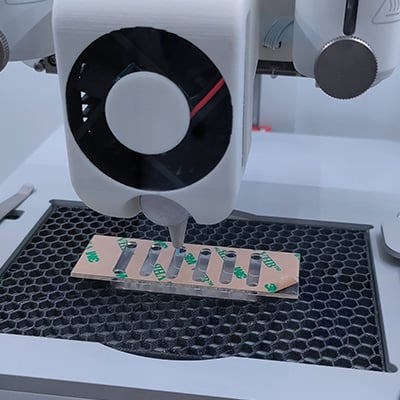
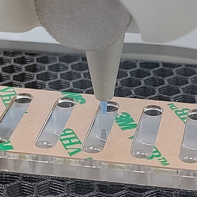
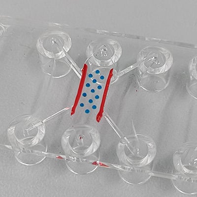
What’s an Easy Way to Seal and Perfuse 3D Bioprinted Structures?
I use the sticky-Slide VI 0.4 to print structures directly into the open channels using a 3D bioprinter. Once the structures are in place, I can easily attach a coverslip to the sticky side of the slide, forming a closed, perfusable system. This method enables me to combine precise 3D bioprinting with in vivo-like cultivation conditions for the printed constructs.

Philipp, R&D Engineer
As a physicist with a focus on biophysics, I am highly interested in 3D printing, stem cells, microfluidics, and organ-on-chip technologies.
How Do I Reduce Evaporation Effects During Imaging in the µ-Slide 15 Well 3D?
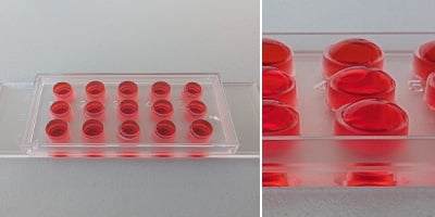
I slightly overfill the wells of the µ-Slide 15 Well 3D so that the medium touches the lid. This simple trick creates ideal conditions for phase contrast microscopy—even when some evaporation occurs. It was a real game changer in my experiments, which used phase contrast microscopy to study the effects of drugs on cardiomyocyte contractility across various gel matrices.

Julia, Technical Marketing Specialist
After more than seven years of working in research at ibidi, I now enjoy sharing my knowledge with ibidi customers through technical marketing.
What’s the Best Way to Test Gels Before Using the µ-Slide I Luer 3D?
Before jumping into your main experiments in the µ-Slide I Luer 3D (e.g., transmigration studies), I always recommend running a test series with different gels and various concentrations using the µ-Slide 15 Well 3D or even the µ-Plate 96 Well 3D. This helps you figure out in advance which gel matrix and concentration work best for your specific cell type and experimental setup, saving money, time, and resources.

Charlotte, Strategic Account Manager
Over the past eight years of working with ibidi users and a wide range of applications, I’ve gathered and shared many useful tips and tricks.
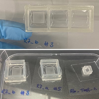
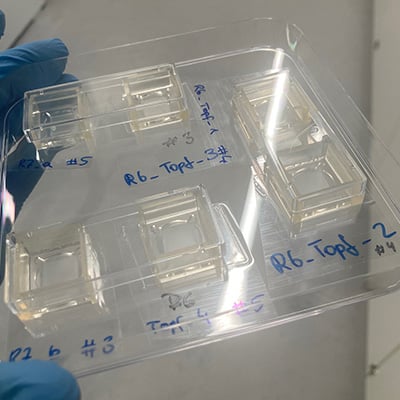
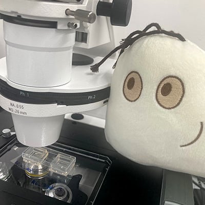
How Do I Create Temporary Culture Chambers on Any Substrate?
I use ibidi silicone Culture-Inserts and Removable Chambers to create temporary, perfectly sized culture chambers on any substrate. The silicone can be cut to fit and adheres without the need for additional glue. With these flexible instant wells, I can perform precise, spatially controlled coating, culturing, and staining while using only a small amount of reagent. Afterwards, the chambers can be removed without leaving any residue, preserving the sample for analysis. This simple technique makes cell culture on diverse materials easy and reliable.

Lea, Senior Research Scientist
I work closely with academic and research partners on exploring new frontiers in cell-based assays.
What Are the Benefits of Using Channel Slides for Fluorescence Staining?
My tip is performing fluorescence staining in the µ-Slide VI 0.4. Many scientists immediately associate channels with flow experiments and don’t consider using them for simple static applications. However, there’s no meniscus effect, the cells distribute evenly, and you only need 30 µl of antibody solution to fill the channel—saving money, especially when working with expensive antibodies.

Carla, Life Science Sales & Application
I worked in research for many years and now enjoy sharing my passion for microscopy with my customers.
Got More Tips & Tricks?
We'd love to hear how you're using ibidi products! Send us your images or videos and a short description to info@ibidi.de.
Thanks for supporting the scientific community by sharing your Tips & Tricks.


