


CellArt Gallery
Image of the Month
2026 ibidi Calendar, July
Wendy Stam
IBL, Leiden University, the Netherlands
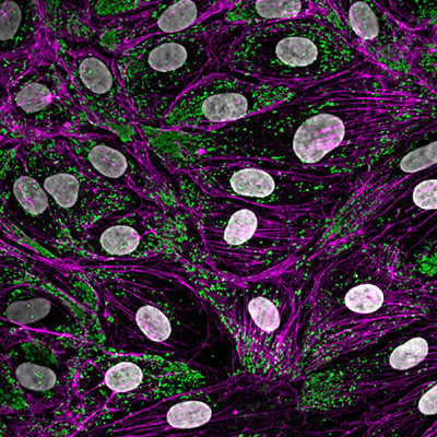
Human umbilical vein endothelial cells (HUVEC) were cultured in a μ-Slide 8 Well high with a gelatin coating. The cells were stained for von Willebrand factor (DAKO, green), actin cytoskeleton (phalloidin, magenta) and nuclei (Hoechst, white). The image is a maximum intensity projection (z = 2 μm, step size = 0.5 μm) taken with a 63x oil objective on a Leica Stellaris 5 microscope.
Follow Wendy Stam on LinkedIn.
List of pages in %s: