


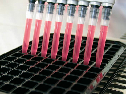
High-Throughput and High-Content Screening
Multiwell plates are the gold standard for high-throughput screening (HTS) and high content screening (HCS) in cell culture. The ibidi µ-Plates provide essential features that make them ideal for HTS/HCS when using high-resolution microscopy and fluorescence-based imaging:
- Compatible with robotics and plate readers due to an ANSI/SLAS (SBS) standard format (85.5 x 127.5 mm)
- Available with a #1.5 Polymer Coverslip bottom with extremely low autofluorescence for undisturbed fluorescence microscopy and ibiTreat (tissue culture-treated) surface for optimal cell attachment
- Available with a #1.5H Glass Coverslip bottom with highest optical quality for special microscopic applications (e.g., TIRF, super-resolution microscopy)
- Compatible with solvents for staining and fixation, as well as immersion oil
- Excellent inner well and whole plate flatness
HTS Application Examples for ibidi µ-Plates
- Compound toxicology screenings and drug screenings
- Large-scale transfection experiments
- Large-scale fluorescence microscopy
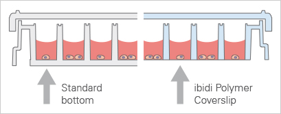
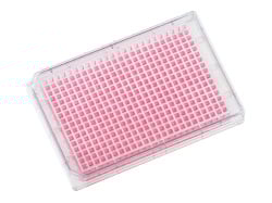
Standard 96 Well Plate With a 1 mm thick bottom made of polystyrene, not suited for high-resolution or fluorescence microscopy. | ibidi µ-Plate 96 Well With a flat ibidi Polymer Coverslip #1.5 bottom (180 µm, +10/–5 µm), ideal for high-resolution or fluorescence microscopy. |
ibidi Blog Article |
Check out the ibidi Blog article High-Content Screening: From Microscopy to Medicine to learn more about the diverse applications of High-Content Screening.
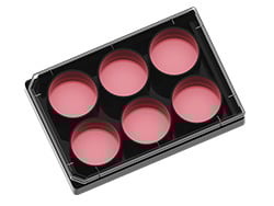
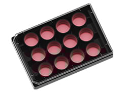
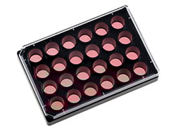
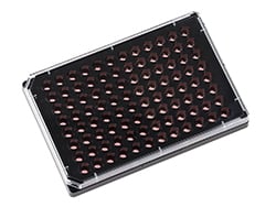
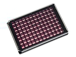
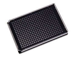
The ibidi µ-Plates Variety
Want to know if you should use a glass or a polymer bottom for your application? Visit our Surface Material Guide. |
High-Throughput Live Cell Imaging Solutions
Live cell imaging is the time-lapse microscopy of dynamic processes in living cells. Several imaging techniques can be applied, such as phase contrast microscopy or confocal microscopy. During the whole live cell imaging experiment, the cells need to be kept alive and healthy. Therefore, physiological conditions have to be established and maintained on the microscope.
ibidi offers Stage Top Incubators for large-scale live cell imaging using microplates that have an ANSI/SLAS (SBS) standard format.
Find Out More
Please find more detailed information about the planning, conduction, and data analysis of live cell imaging assays in the "Live Cell Imaging" application chapter.
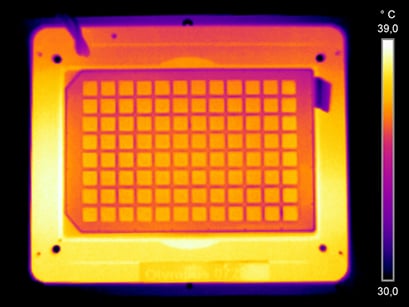
Stable and consistent temperature distribution in every well of multiwell plates in the ibidi Stage Top Incubators. Images were acquired with a FLIR thermal camera.
ibidi SolutionsThe ibidi Stage Top Incubators allow for easy live cell imaging on every inverted microscope, providing precise control of temperature, humidity, and CO2 (optionally O2). They are compatible with all inverted microscopes that have a K-Frame stage (160 mm x 110 mm) or a Nikon Ti-S-E and Ti-S-ER Motorized Stage. |
|
High-Throughput Wound Healing Assays
Wound healing and migration assays are widely used approaches for the analysis of cell migration under different conditions (e.g., when performing drug screenings).
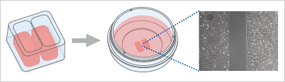
With the Culture-Inserts, ibidi offers a solution for reproducible large-scale wound healing and migration experiments, requiring only a few steps from sample preparation to image analysis:
- Cells are seeded in the single wells, where they attach
- Removal of the silicone insert results in two precisely defined cell patches, which are separated by a 500 µm gap
- Wound closure can now be monitored by using live cell imaging or by taking photos at different time points
Find Out More
Please find more detailed information about the planning, conduction, and data analysis of wound healing and migration assays in the "Wound Healing and Migration" application chapter.
ibidi SolutionsThe ready-to-use Culture-Insert 2 Well 24 is ideal for reproducible high-throughput wound healing and migration assays. It consists of silicone Culture-Inserts with a defined cell-free gap that are already inserted into the µ-Plate 24 Well. Find a PDF of the detailed protocol for performing a wound healing assay using the Culture-Insert 2 Well 24 in the ibidi Application Note "Wound Healing Assay with the Culture-Insert 2 Well 24" (AN 36). |
|
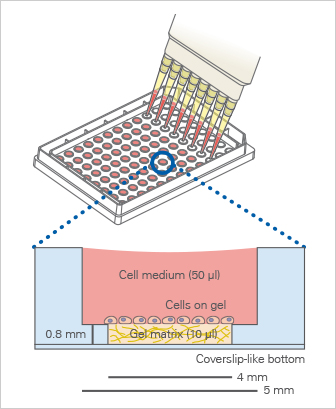
High-Throughput Tube Formation Assays
Tube formation assays are a widely used in vitro tool for accessing angiogenesis in an easy, cost-effective, and reproducible manner. A tube formation assay is performed by first seeding single cells, and then observing and imaging the tube formation over time. From this, several readouts can be analyzed over time, such as tube length and the number of loops.
With the µ-Plate 96 Well 3D, ibidi offers a solution for tube formation experiments with only a few steps from sample preparation to image analysis. Due to the “well-in-a-well” technology, the amount of gel needed is reduced to only 10 μl per well. In addition, no meniscus is formed, ensuring the formation of a uniformly thick gel matrix.
For scientists who need only a small number of samples, ibidi also offers the µ-Slide 15 Well 3D with 15 wells.
Find Out More
Please find more detailed information about the planning, conduction, and data analysis of angiogenesis assays in the "Angiogenesis" application chapter.
ibidi SolutionsThe µ-Plate 96 Well 3D is a multiwell plate with full ANSI/SLAS (SBS) and robotics compatibility. Due to the well-in-a-well principle, it is ideal for cost-effective tube formation experiments, requiring only 10 µl of gel per well. In addition, it allows for brilliant cell visualization without meniscus formation. Find practical advice on how to run tube formation assays using the µ-Plate Angiogenesis 96 Well in the ibidi Application Note “Tube Formation in µ-Plate 96 Well 3D” (AN 05) as a PDF. |
|
Selected Publications for High-Throughput Screening Assays
The pro-angiogenic activity of periodontal ligament stem cells and dental pulp stem cells was quantified using the µ-Plate 96 Well 3D for standardized high-throughput tubulogenesis assays on growth factor-reduced Matrigel.
Roato I, Orrico C, Meinardi S, Pedraza R, Mosca Balma A, Baima G, Genova T, Aimetti M, Mussano F. The Pro-Angiogenic Potential of Periodontal Ligament Stem Cells and Dental Pulp Stem Cells: A Comparative Analysis. Cells. 2025;14(12):864. doi:10.3390/cells14120864.
Read article
High-throughput drug testing of 3D bioprinted triple-negative breast cancer stem cell tumor-stroma models was performed using the µ-Plate 96 Well Square for CSC-targeted therapy evaluation.
González-Callejo P, García-Astrain C, Herrero-Ruiz A, Henriksen-Lacey M, Seras-Franzoso J, Abasolo I, Liz-Marzán LM. 3D Bioprinted Tumor-Stroma Models of Triple-Negative Breast Cancer Stem Cells for Preclinical Targeted Therapy Evaluation. ACS Appl Mater Interfaces. 2024;16(21):27151–27163. doi:10.1021/acsami.4c04135.
Read article
High-content imaging-based phenotypic drug screening in zebrafish xenografts was established in 96-well format using the µ-Plate 96 Well Square for automated microscopy workflows.
Sturtzel C, Grissenberger S, Bozatzi P, et al. Refined high-content imaging-based phenotypic drug screening in zebrafish xenografts. npj Precision Oncology. 2023;7:44. doi:10.1038/s41698-023-00386-9.
Read article
A multilayer 3D cervical cancer model for high-throughput drug screening was established using the µ-Plate 96 Well 3D to study tumor invasion and endothelial microvessel formation.
Cadena IA, Buchanan MR, Harris CG, Jenne MA, Rochefort WE, Nelson D, Fogg KC. Engineering high throughput screening platforms of cervical cancer. J Biomed Mater Res A. 2023. doi:10.1002/jbm.a.37522.
Read article
High-throughput phenotyping of iPSC-derived cardiomyocytes and neurons was performed using the µ-Plate 96 Well Round for fluorescence imaging-based electrophysiology assays and compound testing.
Daily NJ, Du ZW, Wakatsuki T. High-Throughput Phenotyping of Human Induced Pluripotent Stem Cell-Derived Cardiomyocytes and Neurons Using Electric Field Stimulation and High-Speed Fluorescence Imaging. Assay Drug Dev Technol. 2017;15(4):178–187. doi:10.1089/adt.2017.781.
Read article
Find more publications in the ibidi Reference Database.
Frequently Asked Questions
What is high-throughput screening (HTS) and how does it differ from high-content screening (HCS)?
High-throughput screening (HTS) refers to the rapid analysis of large numbers of samples using automated workflows, typically generating simple quantitative readouts such as fluorescence intensity or viability. High-content screening (HCS) extends HTS by incorporating microscopy and image-based analysis, enabling multiparametric evaluation of cellular morphology, protein localization, and dynamic processes in in vitro systems.
Why are ibidi µ-Plates suitable for high-resolution HTS applications?
ibidi µ-Plates feature a #1.5 polymer or #1.5H glass coverslip bottom with excellent optical quality, enabling high-resolution fluorescence and microscopy-based readouts. Their ANSI/SLAS-compatible format ensures seamless integration with robotic systems and plate readers, while low autofluorescence and high plate flatness improve data quality and reproducibility. Examples include the µ-Plate 96 Well 3D, µ-Plate 96 Well Round, and µ-Plate 384 Well Glass Bottom.
Why is inner well flatness important in high-throughput screening?
Inner well flatness ensures that cells grow on a uniform surface and remain within the same focal plane during imaging. This is particularly important for automated microscopy and high-content screening, where even small variations can affect focus, image quality, and quantitative readouts. High inner well flatness helps reduce variability between wells and improves the reliability and comparability of screening results across entire plates.
How can live cell imaging be implemented in high-throughput screening workflows?
High-throughput live cell imaging requires stable environmental conditions across all wells. The ibidi Stage Top Incubators maintains temperature, CO2, and humidity directly on the microscope, allowing continuous time-lapse imaging of cellular processes such as migration, proliferation, and signaling across entire multiwell plates.
What are the advantages of black-walled plates for fluorescence-based assays?
Black-walled plates reduce well-to-well crosstalk and minimize background fluorescence, which is critical for sensitive fluorescence-based assays. Combined with optically clear bottoms, this design improves signal-to-noise ratio and enables accurate quantification in high-content imaging.
How can wound healing assays be adapted for high-throughput screening?
High-throughput wound healing assays can be performed using the Culture-Insert 2 Well 24, which generates defined 500 µm gaps directly in a multiwell plate format. This allows parallelized migration experiments with high reproducibility and compatibility with automated imaging workflows.
How can angiogenesis assays be scaled for high-throughput applications?
Angiogenesis assays can be adapted to high-throughput formats using the µ-Plate 96 Well 3D, which enables parallel tube formation experiments with minimal gel volumes. The well-in-a-well design ensures uniform matrix thickness and reproducible assay conditions across multiple wells.
Which parameters are typically analyzed in high-content screening assays?
Common parameters include cell viability, morphology, proliferation, migration, fluorescence intensity, protein localization, and network formation. Automated image analysis allows extraction of multiple quantitative features per well, enabling comprehensive evaluation of cellular responses across large datasets.