


Angiogenesis
Angiogenesis assays are essential tools to study how new blood vessels form from existing vasculature. This highly regulated process plays a central role in embryonic development, tissue regeneration, wound healing, and reproductive biology. At the same time, dysregulated angiogenesis contributes to major diseases including cancer, cardiovascular disorders, chronic inflammation, and ischemic injury.
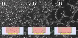
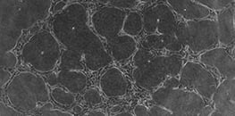
In vitro angiogenesis models allow researchers to recreate key steps of vascular growth under controlled conditions. Typical assays examine endothelial cell migration, alignment, sprouting, and the formation of capillary-like networks in defined extracellular matrix environments. These systems help quantify pro- and anti-angiogenic effects of drugs, growth factors, genetic modifications, and environmental stimuli such as hypoxia.
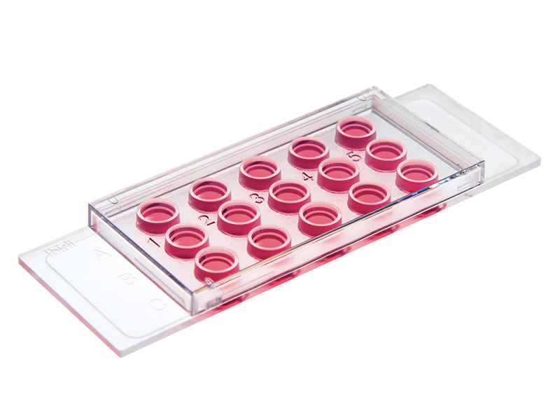
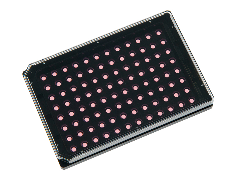
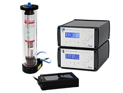
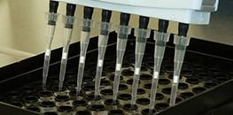
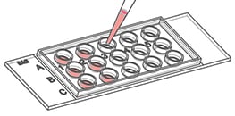
ibidi supports angiogenesis research with imaging-optimized 3D labware and live cell microscopy solutions. The µ-Slide 15 Well 3D and µ-Plate 96 Well 3D provide standardized gel geometry for reproducible tube formation and sprouting assays while minimizing matrix consumption. For long-term and time-lapse experiments, the ibidi Stage Top Incubators maintains stable physiological conditions directly on the microscope, enabling high-quality live cell imaging throughout the entire assay.
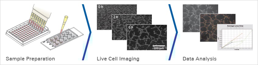
Experimental Workflow for Angiogenesis Assays
ibidi Products for Angiogenesis Assays
Find more products for this application here.
Selected Publications for Angiogenesis Assays
The role of IGF-2 and its variants in endothelial tube formation was analyzed using the µ-Slide 15 Well 3D for standardized Matrigel-based angiogenesis assays.
Alders L, Pirlet E, Gesquiere E, Bronckaers A. The role of IGF-2 and its variants in enhancing endothelial migration and angiogenesis. Front Cell Dev Biol. 2025;13. doi:10.3389/fcell.2025.1598705.
Read article
The pro-angiogenic activity of periodontal ligament stem cells and dental pulp stem cells was quantified using the µ-Plate 96 Well 3D for standardized tubulogenesis assays on growth factor-reduced Matrigel.
Roato I, Orrico C, Meinardi S, Pedraza R, Mosca Balma A, Baima G, Genova T, Aimetti M, Mussano F. The Pro-Angiogenic Potential of Periodontal Ligament Stem Cells and Dental Pulp Stem Cells: A Comparative Analysis. Cells. 2025;14(12):864. doi:10.3390/cells14120864.
Read article
Anti-angiogenic effects of extracellular vimentin targeting were quantified using the µ-Slide 15 Well 3D in Matrigel-based endothelial tube formation assays.
van Beijnum JR, Huijbers EJM, van Loon K, Blanas A, Akbari P, Roos A, Wong TJ, Denisov SS, Hackeng TM, Jimenez CR, Nowak-Sliwinska P, Griffioen AW. Extracellular vimentin mimics VEGF and is a target for anti-angiogenic immunotherapy. Nat Commun. 2022;13:2842. doi:10.1038/s41467-022-30063-7.
Read article
VEGF-A-mediated endothelial angiogenesis and cytoskeletal dynamics were investigated using Matrigel tube formation assays in the µ-Slide 15 Well 3D and live cell imaging workflows.
Luo J, Lu C, Chen Y, et al. Nuclear translocation of cGAS orchestrates VEGF-A-mediated angiogenesis. Cell Rep. 2023;42(4):112328. doi:10.1016/j.celrep.2023.112328.
Read article
Find more publications in the ibidi Reference Database.
Application Notes for Angiogenesis Assays
Videos for Angiogenesis Assays


Frequently Asked Questions
What is angiogenesis and why is it important to study?
Angiogenesis is the process by which new blood vessels grow from pre-existing vasculature. It is essential for embryonic development, tissue regeneration, wound healing, and physiological remodeling. Dysregulated angiogenesis is a hallmark of many diseases, including cancer, cardiovascular disorders, chronic inflammation, and ischemic injury. Studying angiogenesis helps researchers understand disease mechanisms and develop targeted therapeutic strategies using standardized in vitro and in vivo models.
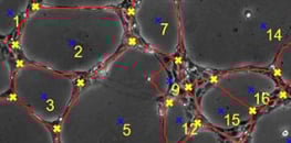
What is a tube formation assay and what does it measure?
A tube formation assay is a widely used in vitro method to model angiogenesis. Endothelial cells are seeded onto a basement membrane-like matrix where they attach, migrate, align, and form capillary-like networks. Quantitative readouts include total tube length, branching points, loop formation, and network area, enabling evaluation of pro- and anti-angiogenic effects under defined experimental conditions.
Why are µ-Slide 15 Well 3D and µ-Plate 96 Well 3D ideal for tube formation assays?
The µ-Slide 15 Well 3D and µ-Plate 96 Well 3D feature a well-in-a-well geometry that creates a uniform gel layer without gel meniscus formation. This ensures consistent matrix thickness and keeps all vascular structures within a single focal plane for microscopy. The design reduces matrix consumption to as little as 10 µl per well while enabling highly reproducible assay conditions.
Which extracellular matrices can be used for angiogenesis assays?
Endothelial cells require a basement membrane-like matrix to form vascular structures. Common options include Matrigel®, collagen gels, laminin-rich matrices, fibrin gels, and synthetic hydrogels. Matrix composition strongly influences tube formation dynamics, structural stability, and morphology, making matrix selection a critical experimental parameter. To reduce reagent costs, low-volume systems like the µ-Slide 15 Well 3D limit gel requirements to 10 µl per well and ensure consistent experimental conditions.
What is the difference between tube formation and sprouting assays?
Tube formation assays analyze how individual endothelial cells organize into capillary-like networks on a matrix surface. Sprouting assays, in contrast, use multicellular spheroids or tissue explants embedded within a 3D gel to evaluate radial vessel outgrowth and invasion into the surrounding matrix. Sprouting assays better mimic 3D vascular invasion, while tube formation assays provide faster and more standardized quantification.
How can angiogenesis assays be combined with live cell imaging?
Angiogenesis is a highly dynamic process unfolding over several hours. Using the ibidi Stage Top Incubators, cells can be maintained under stable temperature, CO2, and humidity directly on the microscope. This enables continuous time-lapse imaging of tube formation, sprouting dynamics, and vascular network maturation without disturbing the culture environment. Combining live imaging with the µ-Plate 96 Well 3D allows parallelized high-content angiogenesis studies.
Which parameters are typically analyzed in angiogenesis assays?
Common quantitative parameters include total tube length, number of junctions, loop count, sprout length, network area, and branching complexity. These metrics can be assessed manually or using automated image analysis tools. Statistical comparison across experimental conditions reveals pro- or anti-angiogenic effects of compounds, genetic modifications, or environmental stimuli.
List of pages in %s: