Microscopy Parameters of Materials
Find Out More
Contact us for a printed version or download the posters "Microscopy With ibidi" or "Choosing the Best Microscopy Labware" as PDF.
The thickness of the coverslip is a crucial parameter that defines imaging quality. Most of the objective lenses used for microscopy are corrected to the standard coverslip thickness of 0.17 mm (170 µm +20/–10 µm, #1.5). Thinner or thicker coverslips require the use of the correction collar on the objective lense, which then prevents the formation of blurred images by spherical and chromatic aberrations.
The ibidi Polymer Coverslip is the standard bottom of all ibidi μ-Slides, μ-Dishes, and μ-Plates. With a thickness of 180 µm (+10/–5 µm), the ibidi Polymer Coverslip provides ideal prerequisites for brilliant inverse high-resolution microscopy.
For special microscopy applications, the μ-Dish 35 mm, high, the µ-Slide 2 Well | 4 Well | 8 Well, and the µ-Slide 2 Well Ph+ | 4 Well Ph+ are also available with a 170 µm (+/–5 µm) coverslip glass bottom #1.5H.
Find more information about the bottom material compatibilities with different microscopic techniques in the chapter, “Comparison of Material Specifications”.
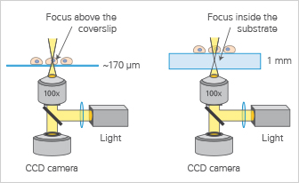
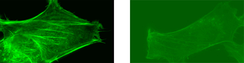
Microscopic lenses are optimized for the standard coverslip thickness of 170 µm (left). Using other coverslip thicknesses makes it impossible for high resolution objective lenses to focus on the specimen (right).
The refractive index nD measures the speed of light inside a specific material, as compared to the absolute vacuum. It is an important value for calculating the numerical aperture (NA).
The refractive index is defined by: nD = c/η
- c = velocity of light in free space
- η = light velocity in a particular medium
The refractive index is wavelength-dependent. As a standard, nD is used to characterize the refractive index of optical materials, which represents 589 nm.
The refractive index is often referred to as “optical density”. Most objectives are designed for use with coverslips that have a standard refractive index of 1.52, including glass and the ibidi Polymer Coverslip. Immersion oil, which can be seen as an extended front lens of immersion objectives, also has a refractive index of 1.52.
| Material | Refractive Index nD |
| Vacuum | 1 (by definition) |
| Air | 1.0003 |
| Water | 1.33 |
| Glycerol | 1.47 |
| Immersion oil | 1.52 |
| Glass coverslip | 1.52 |
| ibidi Polymer Coverslip *includes ibiTreat, Untreated and Bioinert | 1.52 |
The numerical aperture (NA) is an important value for microscope objectives, which defines their resolution and luminous intensity. It measures the ability of the objective to gather light and resolve fine specimen detail at a fixed object distance. The higher the NA, the greater the ability of an objective is to resolve details of a specimen. The NA is imprinted on every objective.
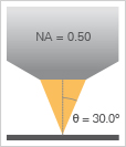
Example of a 20x objective lens with a numerical aperture of 0.5 (20x/0.5).
The numerical aperture is defined by: NA = n sin θ.
- n = refractive index of the medium in which the lens works (e.g., 1.52 for immersion oil)
- θ = half-angle of the maximum cone of light that can enter into the lens
For dry objectives, the maximal NA is ~0.95. For immersion objectives, the maximal NA is ~1.4.
Identical Magnification—Different Optical Resolution
| Low NA Lens Objective: 10x, NA: 0.25 | High NA Lens Objective 10x, NA: 0.45 |
Highly magnified image section to visualize the different resolution
Material Dispersion / Abbe Number
Material dispersion is defined as a variation in the refractive index that depends on the wavelength. In other words, dispersion is a measurement for chromatic aberrations.
Dispersion in an optical material is quantified using the Abbe number. It is calculated from the refractive indices of three different wavelengths. Using a material with a high Abbe number means that the refraction index of different wavelengths is nearly equal in that material, leading to reduced separation of different wavelengths. Therefore, materials with a higher Abbe number give less color dispersion and provide a better optical quality for microscopy.
A material with an Abbe number equal to or larger than 55 is considered to be well-suited for high-resolution microscopy. For example, the ibidi Polymer Coverslip has an Abbe number of 56 and the D 263 M Schott borosilicate glass has an Abbe number of 55.
The transmission of bottom material is an important parameter in microscopy. It describes the material’s ability to permit the passage of light through it at specific wavelengths. The more that light is absorbed by the material, the less it can contribute to fluorescence excitation and image acquisition. To be suitable for different microscopy applications, a material should have a high transmission ability across various wavelengths, such as the ibidi Polymer Coverslip. Unlike standard polystyrene (PS) cell culture surfaces that block the UV light, the ibidi Polymer Coverslip permits UV light transmission at wavelengths below 300 nm.
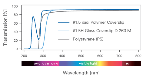
The slide body of the ibidi channel slides and Ph+ µ-Slides is made from the very same plastic microscopy material as the ibidi Polymer Coverslip. Therefore, the transmission properties are identical for the ibidi open wells, channel slides, and Ph+ µ-Slides.
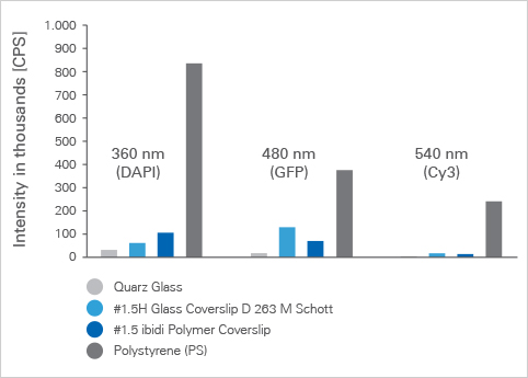
Autofluorescence describes the intrinsic fluorescence intensity of the pure material (e.g., the ibidi Polymer Coverslip or a glass coverslip) without any fluorescent sample. The autofluorescence is emitted by the material and can emerge as noise or background during the imaging process. Autofluorescence can be disruptive, especially when trying to image faint fluorescent signals. All materials and culture media show some degree of autofluorescence, which varies with the excitation/emission wavelength and strongly depends on the material type.
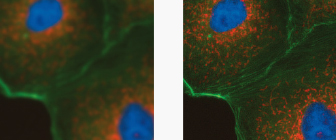
Autofluorescence Influences the Signal-to-Noise Ratio
| Low autofluorescence using the ibidi Polymer Coverslip. | High autofluorescence using a standard culture flask. |
Background Fluorescence of Different Materials for Microscope Slides