


 Micropatterned Surface: Defined Cell Adhesion
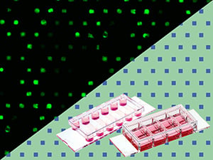
The ibidi µ-Patterning technology enables spatially defined cell adhesion for various 2D and 3D cell culture applications.
Miniaturized adhesive patterns (e.g., lines, squares, or dots) are irreversibly printed on the non-adhesive Bioinert surface of the ibidi Polymer Coverslip, allowing for precisely controlled cell adhesion. The µ-Patterns are dry-stable, sterile, and ready to use.
The µ-Pattern, the Bioinert surface, and the ibidi Polymer Coverslip are all optimized for high resolution imaging and microscopy.
Please find more details about the µ-Patterning technology here.
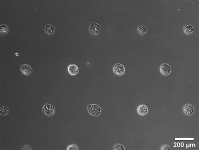
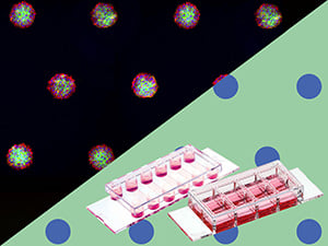
Spheroid formation of NIH-3T3 cells, imaged 14 days after seeding single cells in the µ-Slide VI 0.4 with a multi-cell µ-Pattern. Phase contrast microscopy, 4x objective lens.
The ibiTreat µ-Pattern Variety
Understanding the Key Elements of µ-Patterns
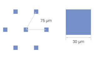
Every ibidi µ-Pattern is designed with precise spatial control, ensuring reproducible cell adhesion and organization. Let's break down the essential pattern components:
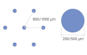
- Shape – Defines the geometry of the attachment area (e.g., cir = circle)
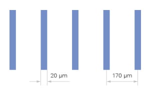
- Size – The diameter or width of the attachment area, measured in micrometers (µm) (e.g., 500 µm)
- Pitch – The spacing between adjacent shapes, measured from center to center (e.g., pit1000 = 1000 µm)
- Layout – The overall pattern arrangement, determining how attachment areas are structured (e.g., a hexagonal layout)
- Surface – The cell attachment surface where cells adhere, made of ibiTreat for optimal cell growth
Looking for other pattern options? Make a special request here, and we will develop a solution tailored to your specific applications.