Top sellers
MV 20: The ibidi Pump System
MV 21: Long-Term Cell Culture Under Perfusion
MV 26: Cell Culture and Microscopy Using the µ-Slide 8 Well
MV 27: Cell Culture and Microscopy Using the µ-Slide I Luer
MV 28: Cell Culture and Microscopy Using the µ-Dish 35 mm, high
MV 32: Tube Formation Assays Using the µ-Slide 15 Well 3D
MV 33: Wound Healing Assays Using the Culture-Insert 2 Well
MV 34: 48 Hours Live Cell Imaging of MDCK Cells
MV 35: Tube Formation Assay of HMEC Cells
MV 37: Chemotaxis Assays Using the µ-Slide Chemotaxis
MV 41: Advanced ibidi Technologies: Micropatterning, 3D Cell Culture and Flow Applications
MV 43: Handling the 8 Well Chamber, removable
MV 46: How to use the µ-Slide Spheroid Perfusion
MV 48: Filling of the µ-Slide I Luer
MV 49: Medium Exchange in the µ-Slide I Luer
MV 50: Filling of the µ-Slide VI 0.4
MV 51: Medium Exchange in the µ-Slide VI 0.4
MV 52: Handling the µ-Slide 8 Well high
MV 53: Handling the µ-Plate 24 Well
MV 54: Handling the µ-Plate 96 Well Square
MV 55: Handling the µ-Plate 96 Well Round
MV 56: Handling the µ-Plate 384 Well Glass Bottom
MV 57: Handling the µ-Slide I Luer 3D
MV 58: Handling the µ-Slide Spheroid Perfusion
MV 59: Handling the µ-Plate 6 Well
MV 60: The Principle of Phase Contrast Microscopy
MV 61: Optimization of Phase Contrast Microscopy
MV 62: The ibidi µ-Patterning Technology
MV 63: Handling the µ-Plate 24 Well for Membrane Inserts
MV 64: Handling the µ-Plate 12 Well
MV 66: The ibidi Micro Illumination System
MV 67: Setup of the ibidi Micro Illumination System
MV 68: Handling of the ibidi Micro Illumination System
MV 69: Handling of the µ-Slide Tissue Engineering
MV 70: Microstructuring with the ibidi Micro Illumination System
MV 71: Using the µ-Slide I Luer 3D
MV 72: Connecting the µ-Slide Spheroid Perfusion to the ibidi Pump System
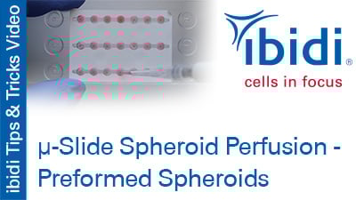
MV 73: Using Preformed Spheroids in the µ-Slide Spheroid Perfusion
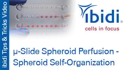
MV 74: Spheroid Self-Organization in the µ-Slide Spheroid Perfusion
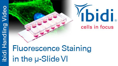
MV 75: Fluorescence Staining in the µ-Slide VI
check_circle