


Immunofluorescence (IF) Application Guide
Immunofluorescence (IF) is a fluorescence microscopy technique used to detect specific proteins or other antigens within cells and tissues using fluorescently labeled antibodies. It plays a central role in cell biology and biomedical research, enabling the visualization of cellular structures, molecular interactions, and signaling pathways with high specificity and resolution.
To perform these experiments, researchers use fluorescent antibody labeling protocols that involve steps such as fixation, permeabilization, blocking, and antibody incubation. These assays are essential for investigating protein localization, expression patterns, and cellular dynamics under normal and pathological conditions.
ibidi supports these applications by offering optimized labware that simplify IF workflows and ensure excellent imaging results. To learn more, explore the subchapters below, which cover key aspects of immunofluorescence, including principles, protocols, troubleshooting tips, and product recommendations.
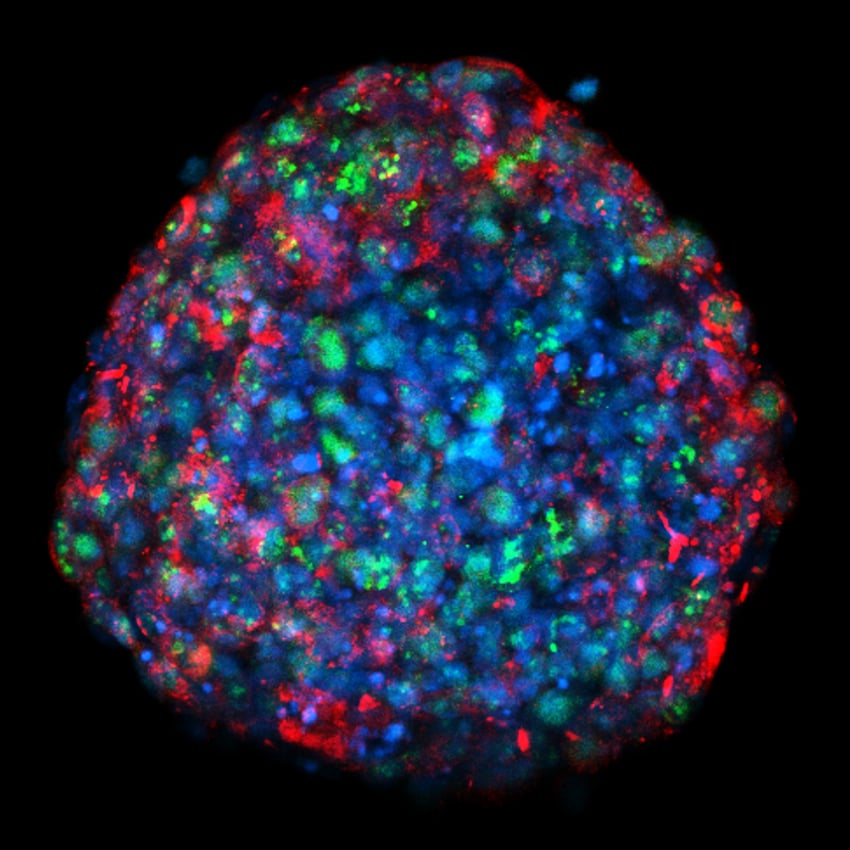
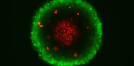
Confocal maximum intensity projection (MIP) image of an 8-day breast tumor spheroid mounted on an ibidi µ-Slide 18 Well Glass Bottom. Ki67+ cells (green), gap junction protein (red), and nuclei (blue). Image by Marina Rodriguez-Candela Mateos, INIBIC, A Coruna, Spain.
Explore the Immunofluorescence Application Guide
Discover the principles, full workflow, recommended controls, and troubleshooting strategies for reliable immunofluorescence staining in cells and tissues. Compare ibidi labware formats to select the best solution for your assay.
ibidi Products for
Immunofluorescence
Choose the optimal products for your immunofluorescence assay.

Carla's Trick:
What are the benefits of using channel slides for fluorescence staining?
Find out on our Tips and Tricks page!
Selected Publications using Immunofluorescence Assays
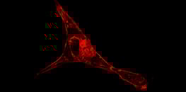
Cancer-induced nerve injury and neurite morphology were analyzed using the µ-Slide 8 Well Glass Bottom for immunofluorescence staining of hPS-cell-derived sensory neurons.
Baruch E.N., Gleber-Netto F.O., Nagarajan P. et al. Cancer-induced nerve injury promotes resistance to anti-PD-1 therapy. Nature. 2025;646:462–473. doi:10.1038/s41586-025-09370-8.
Read article
G-quadruplex DNA structures in human stem cells and differentiation models were visualized using the 3 Well Chamber, removable and the µ-Plate 24 Well for immunofluorescence microscopy.
Zyner K.G., Simeone A., Flynn S.M. et al. G-quadruplex DNA structures in human stem cells and differentiation. Nat Commun. 2022;13:142. doi:10.1038/s41467-021-27719-1.
Read article
Mitochondrial inner membrane dynamics were analyzed by live cell STED nanoscopy using the µ-Dish 35 mm, high Glass Bottom for high-resolution imaging.
Liu T., Stephan T., Chen P. et al. Multi-color live-cell STED nanoscopy of mitochondria with a gentle inner membrane stain. Proc Natl Acad Sci U S A. 2022;119(52):e2215799119. doi:10.1073/pnas.2215799119.
Read article
Early human development and germ cell marker expression were analyzed using the µ-Slide 8 Well for cell culture, fixation, and immunofluorescence staining.
Kobayashi T. et al. Principles of early human development and germ cell program from conserved model systems. Nature. 2017;546:416–420. doi:10.1038/nature22812.
Read article
Find more publications in the ibidi Reference Database.
ibidi Blog Article
Are you struggling to get optimal IF results? Shuntaro Yamada from the University of Bergen explains "5 Crucial Steps for Obtaining a Great Immunofluorescent Cell Image".
Application Notes for Immunofluorescence Assays

Immunofluorescence Assay Videos

User Protocols for Immunofluorescence Assays
Webinars for Immunofluorescence Assays
Frequently Asked Questions
What is immunofluorescence (IF)?
Immunofluorescence (IF) is a microscopy-based technique that uses fluorescently labeled antibodies to detect and visualize specific proteins or antigens within cells and tissues. It allows scientists to study protein localization, cellular structures, and molecular interactions with high specificity and spatial resolution.
What is the difference between direct and indirect immunofluorescence?
In direct immunofluorescence, the primary antibody is directly conjugated to a fluorophore, providing a quick, one-step labeling process. In indirect immunofluorescence, an unlabeled primary antibody binds the target, followed by a fluorophore-labeled secondary antibody for signal amplification. The indirect method is more commonly used for higher sensitivity and flexibility.
How does immunofluorescence (IF) differ from immunohistochemistry (IHC)?
Both techniques use antibodies to detect specific proteins. Immunofluorescence (IF) uses fluorescent dyes and requires a fluorescence microscope, while immunohistochemistry (IHC) uses enzymatic or colorimetric reactions that are visible under a brightfield microscope. IHC is typically used on tissue sections, whereas IF is often applied to cultured cells or thin tissue slices. IF allows for higher spatial resolution and multiplex staining, enabling precise co-localization of multiple targets.
What are the key steps in an immunofluorescence staining protocol?
A typical immunofluorescence staining protocol includes fixation, permeabilization, blocking, incubation with primary and secondary antibodies, and mounting with an antifade medium. Each step is critical to preserve cell morphology, minimize background, and achieve strong, specific fluorescence signals.
How can I improve my immunofluorescence staining results?
Start by optimizing fixation and permeabilization, as these steps are critical for preserving cellular structures and allowing antibody access. Use carefully titrated antibody concentrations and include a blocking step to minimize background staining. Make sure to follow proper incubation times and wash thoroughly between steps. Minimize photobleaching by reducing light exposure and using antifade mounting media. ibidi’s Mounting Medium with DAPI helps preserve fluorescence signals for high-quality imaging results.
Can immunofluorescence be combined with live cell imaging?
Typically, immunofluorescence is performed on fixed cells. However, live cell imaging can be achieved using genetically encoded fluorescent proteins or live cell compatible dyes. ibidi’s Chambered Coverslips and Stage Top Incubators support optimal environmental conditions for live fluorescence microscopy, enabling time-lapse studies and dynamic analysis.
What microscopy techniques can be used with ibidi products for immunofluorescence?
All ibidi Slides, Dishes and Multiwell Plates are designed for compatibility with a wide range of microscopy techniques. These include widefield fluorescence, confocal, spinning disk, TIRF, and super-resolution microscopy. The high-optical-quality #1.5 polymer or #1.5H glass coverslip bottoms ensure optimal imaging performance across standard and advanced fluorescence applications.