


Immunofluorescence Applied: Experimental Examples
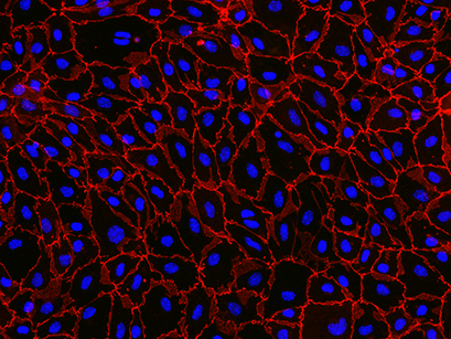
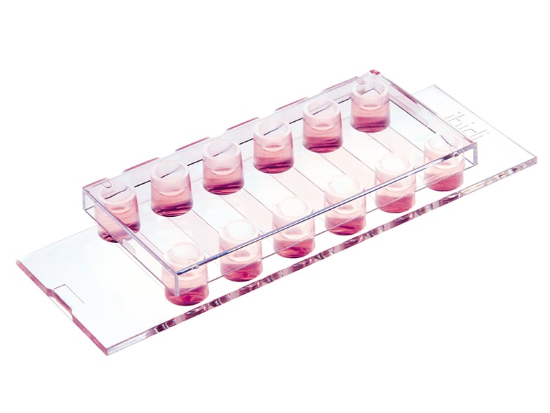
Immunofluorescence staining of primary mouse brain microvascular endothelial cells (pMBMECs), cultured in the ibidi 12 Well Chamber, removable. Red color delineates the endothelial cell junctions labeled for zonula occludens (ZO)-1, highlighting their matured state. Blue color shows the nuclei stained with DAPI. The image was acquired with a 20x objective on a Nikon Eclipse microscope.
Data by Sidar Aydin, Britta Engelhardt, Ruth Lyck, Theodor Kocher Institute, Bern, Switzerland.
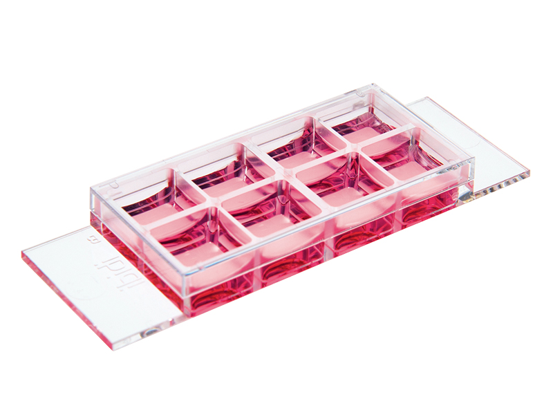
ibidi product used:

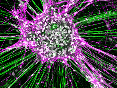
Rat dorsal root ganglionic cells and Schwann cells cultured in an ibidi μ-Slide 8 Well and stained for neurofilament (green), NGFR (magenta), and DAPI (white). The image was obtained with a LEICA SP8X laser scanning microscope.
Data by Tamara Weiss, Division of Plastic and Reconstructive Surgery, Medical University of Vienna, Austria.
ibidi product used:
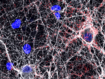
Confocal microscopy of rat hippocampal neurons plated over astrocytes, cultured on ibidi µ-Plate 24 Well. Red is Synapsin 1, a pre-synaptic marker, white is neuron-specific Tubulin Beta-III, and blue is DAPI, a nuclear counterstain.
Data by Daniel Hoeppner, Lieber Institute for Brain Development. Baltimore, MD, USA.
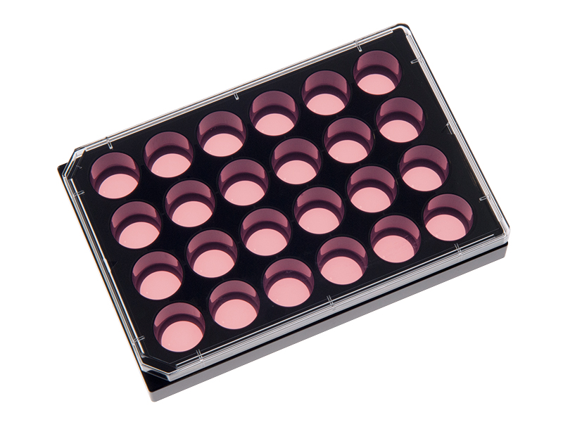
ibidi product used:
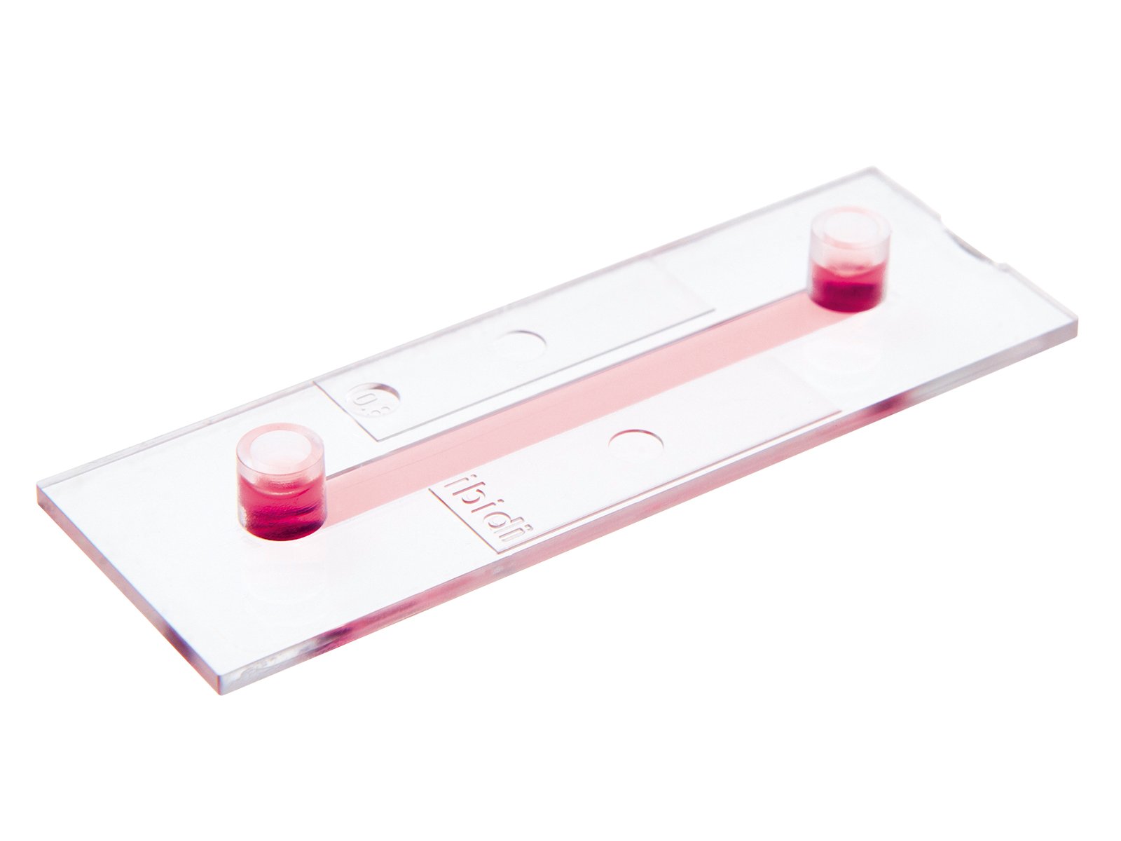
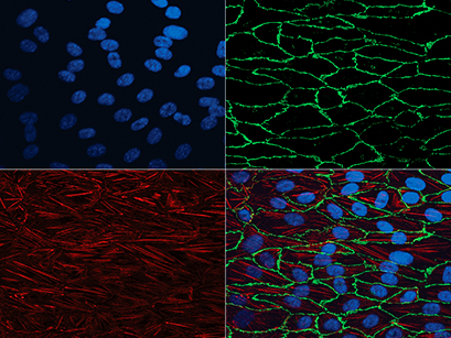
Human umbilical vein endothelial cells (HUVECs) were cultured under flow conditions in the ibidi μ-Slide I 0.4 Luer. Red: actin cytoskeleton (Cy5-conjugated antibody).
Green: adherens junctions, marked by VE-cadherin (Alexa 488-conjugated antibody). Blue: nuclear counterstaining (DAPI).
Data by S. Zahler, Munich, Germany.
ibidi product used:
Madin-Darby canine kidney (MDCK) cells were cultured in the ibidi μ-Slide VI 0.4.
Green: F-actin cytoskeleton (Alexa488-Phalloidin). Red: mitochondria (MitoTracker). Blue: nuclear counterstaining (DAPI). Widefield fluorescence using 100x objective lens with oil immersion.
ibidi product used:

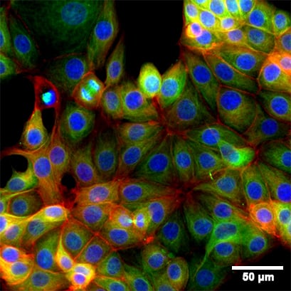
Fluorescence microscopy of immunostained MCF-7 cells in the 8 Well Chamber, removable. Green: alpha-Tubulin. Red: F-actin (phalloidin). Blue: nuclei (DAPI). Widefield fluorescence images were taken with a 20x objective.
ibidi product used:
Read on and learn more about the Principle of Immunofluorescence Assays, or see a typical Workflow of an Immunofluorescence Staining.