



Immunofluorescence Staining Using ibidi Labware
With ibidi labware, all steps of an immunofluorescence staining can be performed in the same microscopy chamber: cell can be cultured, fixed, stained, and imaged directly without any sample or coverslip transfer.
This guide compares the ibidi Chambered Coverslips, Channel Slides, and removable Chamber Slides for immunofluorescence workflows, helping you select the right format based on microscopy setup, reagent volume, handling requirements, and sample storage.
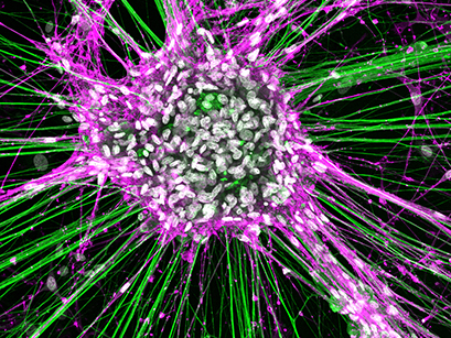
Rat dorsal root ganglionic cells and Schwann cells cultured in a µ-Slide 8 Well and stained for neurofilament (green), NGFR (magenta), and DAPI (white). The image was obtained with a LEICA SP8X laser scanning microscope. Data by Tamara Weiss, Division of Plastic and Reconstructive Surgery, Medical University of Vienna, Austria.
Benefits of ibidi µ-Chambers for Immunofluorescence
- Fast and simple handling
All-in-one chambers simplify your immunofluorescence workflow. - Cost-effective experiments
Requires only a small number of cells and low reagent volumes. - High-resolution imaging
Ideal for many basic and advanced microscopy methods, such as widefield fluorescence, confocal imaging, 2-photon microscopy, and super-resolution microscopy.
ibidi Mounting Medium
High-quality, non-hardening, with or without DAPI, optimized for fluorescence microscopy.
Immunofluorescence Protocol Comparison: Coverslips vs. ibidi Labware
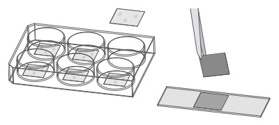
Using ibidi labware significantly shortens the immunofluorescence workflow compared to traditional coverslip methods. Cells can be cultured and stained directly in µ-Slides, µ-Dishes, or µ-Plates, eliminating additional handling steps and reducing protocol time. Explore the step-by-step workflow for traditional immunofluorescence staining using coverslips (left) in comparison to the step-by-step workflow for staining directly in ibidi µ-Slides (right), minimizing handling and reducing overall assay time.
Traditional Immunofluorecence Protocol
With Cells on Coverslip
| Step | Traditional Coverslip Workflow |
|---|---|
| 1 | Sterilize coverslips |
| 2 | Coat coverslips |
| 3 | Place sterile, coated coverslips into 6 well plate |
| 4 | Seed cells in large volume |
| 5 | Wash |
| 6 | Fix → Wash → Permeabilize → Wash → Block |
| 7 | Incubate with primary antibody → Wash → Incubate with secondary antibody → Wash |
| 8 | Peel off the coverslip and transfer to microscopy slide |
| 9 | Mount cells with mounting medium |
| 10 | Seal coverslip with nail polish |
All-In-One Immunofluorescence Protocol
With ibidi µ-Slides
| Step | ibidi µ-Slide Workflow |
|---|---|
| 1 | Skip |
| 2 | Skip |
| 3 | Skip |
| 4 | Seed cells directly in chamber (low volume) |
| 5 | Wash |
| 6 | Fix → Wash → Permeabilize → Wash → Block |
| 7 | Incubate with primary antibody → Wash → Incubate with secondary antibody → Wash |
| 8 | Skip |
| 9 | Mount cells with mounting medium |
| 10 | Skip |
Comparison of ibidi Labware for Immunofluorescence Staining
The table below compares ibidi Chambered Coverslips, Channel Slides, removable Chamber Slides, and traditional 6 well plate workflows for immunofluorescence staining. It summarizes key differences in handling, reagent volume, coverslip requirements, microscopy compatibility, sample storage, and typical use.
| Feature | Chambered Coverslips | Channel Slides | Removable Chamber Slides | Traditional 6 Well Plate |
|---|---|---|---|---|
| Product example | µ-Slide 8 Well high | µ-Slide VI | 8 Well Chamber, removable | Standard 6 well plate (not offered by ibidi) |
| Bottom material | #1.5H Glass Coverslip or #1.5 Polymer Coverslip | #1.5H Glass Coverslip or #1.5 Polymer Coverslip | Standard microscopy glass slide | Polymer |
| IF staining workflow | Seed, stain, and image directly in chambered coverslip | Seed, stain, and image directly in channel slide | Seed and stain on microscopy glass slide, mount with coverslip for imaging | Seed and stain on coverslip, mount on microscopy glass slide for imaging |
| Number of IF protocol steps | Few | Few | Few | Many |
| High-throughput stainings | Limited | Limited | Yes | No |
| Low-volume stainings | Yes | Yes | Limited | No |
| Homogeneous cell & antibody distribution | Limited | Yes | Limited | No |
| Additional coverslips required | No | No | Yes | Yes |
| Typical mounting medium | Non-hardening | Non-hardening | Hardening | Hardening |
| Microscope type | Inverted | Inverted | Inverted & upright | Inverted & upright |
| Sample storage | Short-term | Short-term | Long-term | Long-term |
| Typical use | Parallel immunofluorescence assays without coverslip handling | Low-volume immunofluorescence staining with precise medium exchange | Mounted samples for long-term storage or staining jar workflows | Low-budget IF staining when time is not critical |
The comparison above helps identify the most suitable ibidi labware format for immunofluorescence staining based on sample handling, reagent volume, microscopy method, and storage requirements. Chambered Coverslips and Channel Slides support direct staining and imaging in the same microscopy-ready vessel without handling any additional coverslips, while removable Chamber Slides are better suited for mounted samples and long-term storage. This makes the choice of labware an important factor for reproducible fluorescence staining, reduced sample handling, and efficient immunofluorescence workflows.
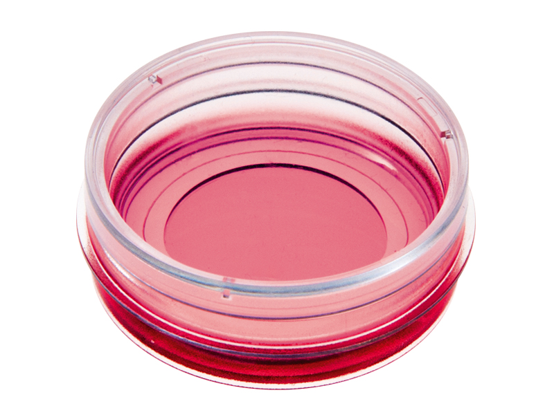
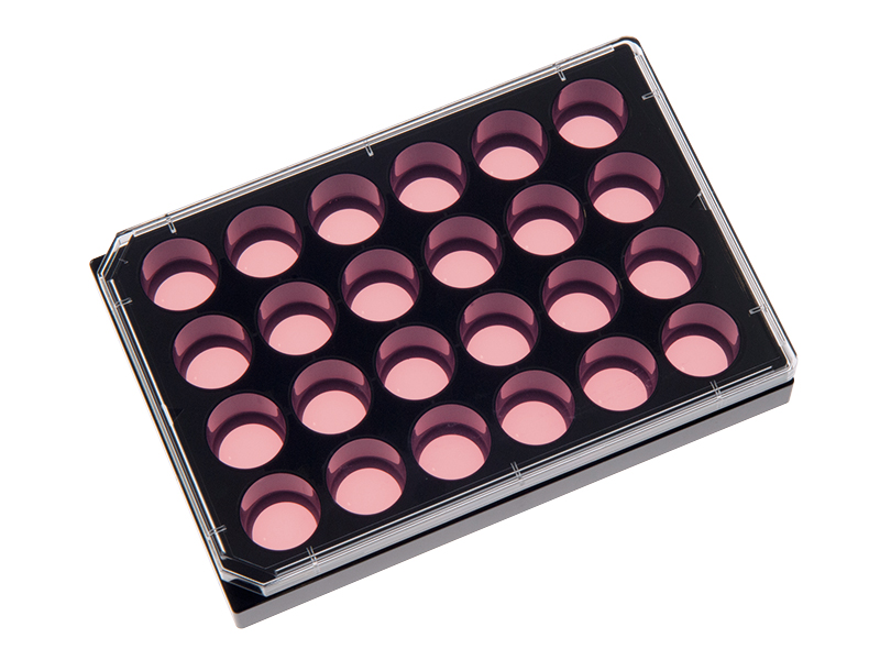
Chambered Coverslips
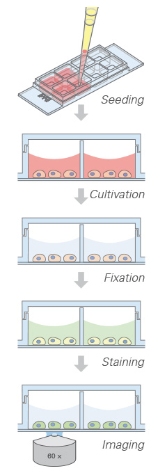
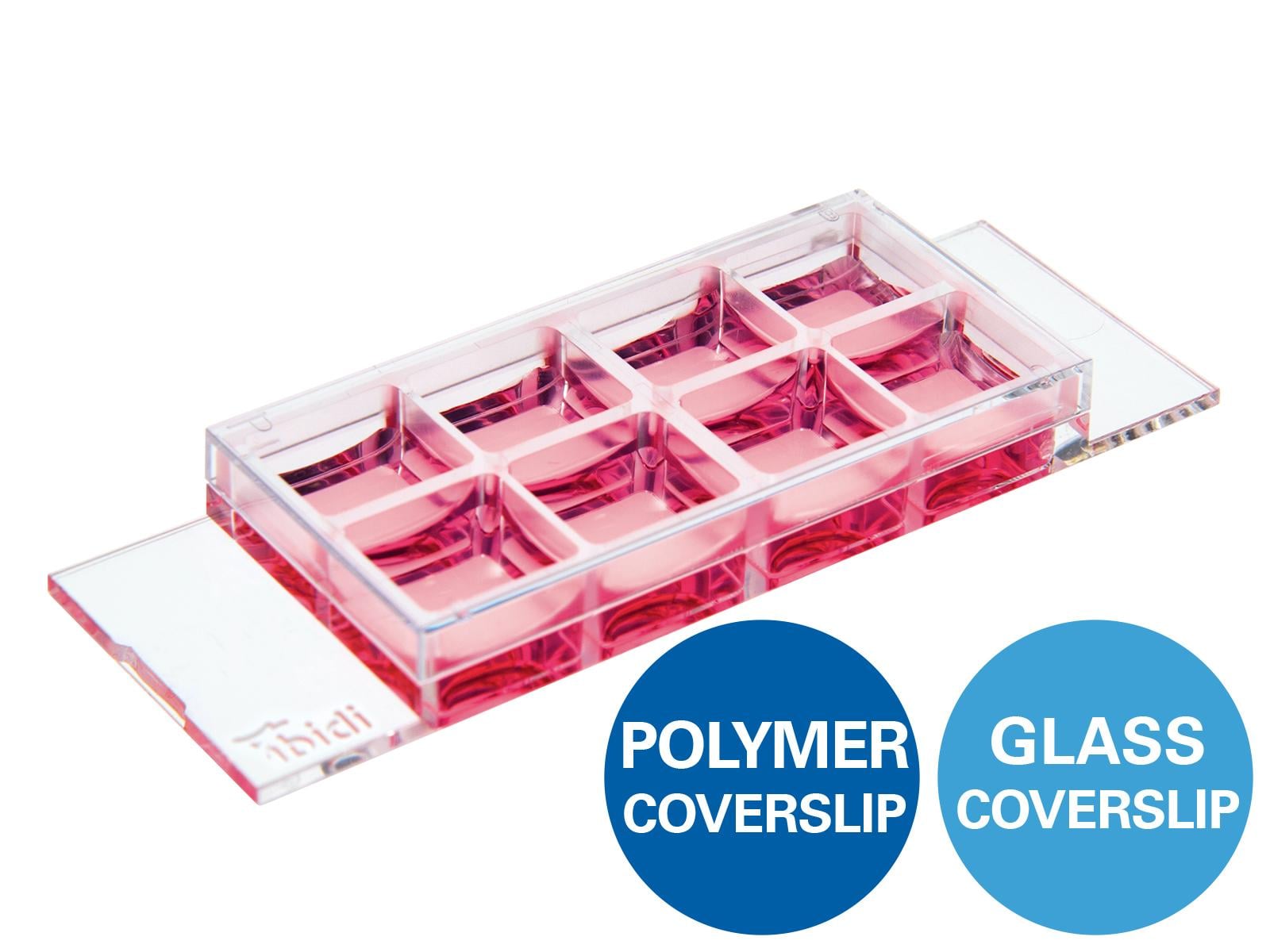
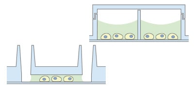
Chambered Coverslips allow performing the entire staining protocol without additional coverslips, with imaging directly through the coverslip bottom.
Advantages
- 1 to 18 non-removable wells on a coverslip bottom
- No coverslip handling
- Parallel assays without cross-contamination
Limitations
- Storage limited to weeks due to gas exchange through polymer
Channel Slides
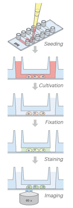
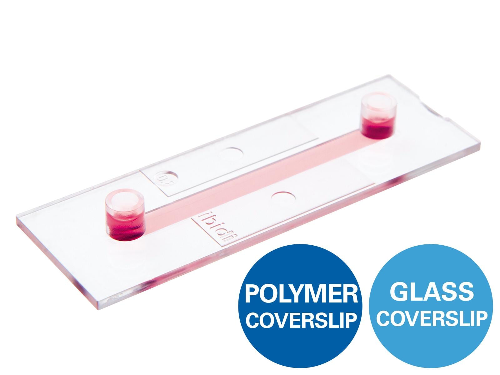
Channel Slides are ideal for low-volume staining with precise medium exchange. The coverslip bottom eliminates the need for additional coverslips.
Advantages
- Many channel heights/coatings
- No coverslip handling
- Low volumes of reagents
- Homogeneous cell and antibody distribution
Limitations
- Storage limited to weeks due to gas exchange through polymer
Removable Chamber Slides
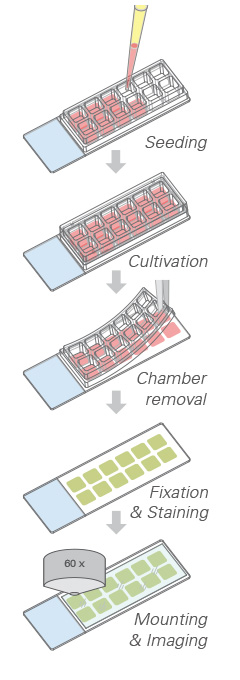
Removable Chamber Slides feature a silicone gasket mounted on a glass slide and are ideal for long-term storage of samples sealed with a coverslip.
Advantages
- Removable silicone chambers on a standard glass slide
- Ideal for long-term storage
- High-throughput screening possible
Limitations
- No high-resolution microscopy during cell cultivation
Immunofluorescence Application Examples in 2D and 3D Cell Culture
These application examples illustrate how ibidi labware supports immunofluorescence staining and fluorescence microscopy across different sample types and assay formats. The selected images show 2D and 3D cell samples, including endothelial junctions, neuronal and glial markers, cytoskeletal structures, mitochondria, and cells cultured under flow conditions.
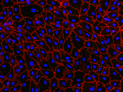
Immunofluorescence staining of primary mouse brain microvascular endothelial cells (pMBMECs), cultured in the 12 Well Chamber, removable. Red: endothelial cell junctions labeled for zonula occludens (ZO)-1. Blue: nuclei stained with DAPI. The image was acquired with a 20× objective on a Nikon Eclipse microscope. Data by Sidar Aydin, Britta Engelhardt, Ruth Lyck, Theodor Kocher Institute, Bern, Switzerland.
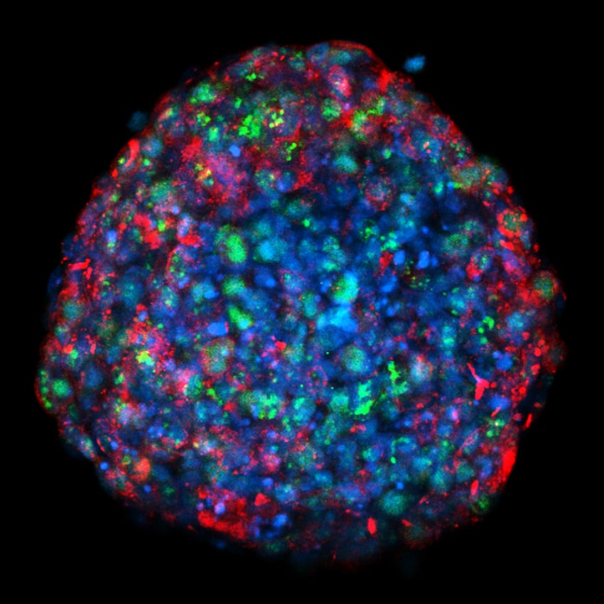
Confocal maximum intensity projection (MIP) image of an 8-day breast tumor spheroid mounted on an ibidi µ-Slide 18 Well Glass Bottom. The image shows stainings for nuclei (blue), Ki67+ cells (green) and gap junction protein (red). Image by Marina Rodriguez-Candela Mateos, Institute of Biomedical Research of A Coruña (INIBIC), A Coruna, Spain.
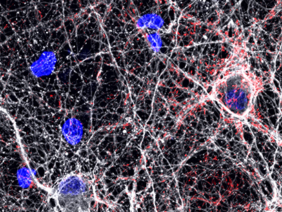
Confocal microscopy of rat hippocampal neurons plated over astrocytes, cultured in a µ-Plate 24 Well. Red: Synapsin 1, a pre-synaptic marker. White: neuron-specific Tubulin Beta-III. Blue: DAPI nuclear counterstain. Data by Daniel Hoeppner, Lieber Institute for Brain Development, Baltimore, MD, USA.
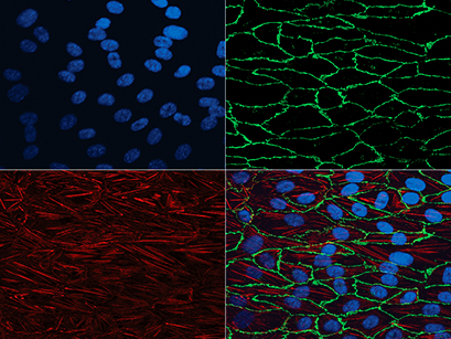
Human umbilical vein endothelial cells (HUVECs) cultured under flow conditions in the µ-Slide I 0.4 Luer. Red: actin cytoskeleton labeled with Cy5-conjugated antibody. Green: adherens junctions marked by VE-cadherin with Alexa 488-conjugated antibody. Blue: nuclear counterstaining with DAPI. Data by S. Zahler, Munich, Germany.
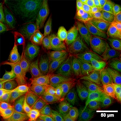
Fluorescence microscopy of immunostained MCF-7 cells in the 8 Well Chamber, removable. Green: alpha-Tubulin. Red: F-actin stained with phalloidin. Blue: nuclei stained with DAPI. Widefield fluorescence images were taken with a 20× objective.
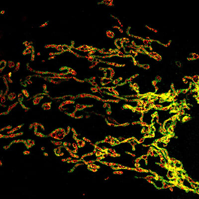
COS7 cells were cultured in the µ-Slide 8 Well high Glass Bottom and immunolabeled for two mitochondrial proteins. Alexa Fluor 594 and Abberior STAR RED signals were imaged by STED microscopy using a 775 nm depletion laser and a 100× oil immersion objective. Image courtesy of Till Stephan, Goethe University, Frankfurt am Main, Germany.
FAQ: Immunofluorescence Staining With ibidi Labware
Which ibidi labware is best for low-volume immunofluorescence staining?
Channel Slides, especially the µ-Slide VI 0.4, are well suited for low-volume immunofluorescence staining. The channel geometry enables precise medium exchange, homogeneous antibody distribution, and reduced reagent consumption during fixation, blocking, antibody incubation, and washing steps.
Can immunofluorescence staining be performed directly in ibidi µ-Slides?
Yes. Cells can be cultured, fixed, permeabilized, blocked, stained, and imaged directly in many ibidi µ-Slides. This reduces coverslip handling, minimizes sample loss, and simplifies the immunofluorescence workflow compared to traditional staining on separate coverslips.
Which ibidi labware is suitable for long-term storage of immunostained samples?
Removable Chamber Slides are suitable for long-term storage of immunostained samples. After staining, the silicone chamber can be removed and the sample can be mounted with hardening mounting medium and a coverslip on a standard glass slide.
Are ibidi Chambered Coverslips compatible with high-resolution fluorescence microscopy?
Yes. ibidi Chambered Coverslips with a #1.5H Glass Coverslip or #1.5 Polymer Coverslip bottom are compatible with high-resolution fluorescence microscopy. They are suitable for common imaging methods such as widefield fluorescence, confocal microscopy, 2-photon microscopy, and super-resolution microscopy (best output with #1.5H Glass Coverslip).
What is the main advantage of ibidi labware compared to traditional coverslip staining?
The main advantage is that cells can be cultivated, stained, and imaged in the same microscopy-ready vessel. This eliminates the need to transfer coverslips, requires fewer handling steps, reduces reagent consumption, and allows immunofluorescence workflows to be performed more quickly and with greater reproducibility.
When should I use Chambered Coverslips instead of Channel Slides?
Chambered Coverslips are useful for parallel immunofluorescence assays, different staining conditions, and easy access to each well. Channel Slides are preferable when very low reagent volumes, precise medium exchange, and homogeneous cell or antibody distribution are critical.
Application Notes and Protocol Resources for Immunofluorescence
- AN 02: Fluorescence Staining using a µ-Slide I (PDF)
- AN 09: Fluorescence Staining using a µ-Slide VI 0.4 (PDF)
- AN 15: Fluorescence Staining using a µ-Slide y-shaped (PDF)
- AN 16: Fluorescence Staining Using the µ-Slide 8 Well high (PDF)
- AN 49: Fluorescence Staining using a 12 Well Chamber, removable (PDF)
- AN 50: Fluorescence Staining using a 3 Well Chamber, removable (PDF)
- AN 45: Mounting Medium Types (PDF)