



Generation and Cultivation of 3D Cell Models
Three-dimensional (3D) cell models can be generated using scaffold-based or scaffold-free culture methods. The most suitable approach depends on the model type, cell source, biological question, required throughput, and downstream analysis. Common 3D cell culture models include spheroids, organoids, embryoid bodies, tissue constructs, and matrix-embedded single cells. These models can also be incorporated into more advanced microphysiological systems (MPS) and organ-on-a-chip platforms that provide controlled flow, mechanical stimulation, or tissue–tissue interactions.
The main methods for generating and cultivating 3D cell models include:
- Cell Cultivation in a 3D Matrix
- 3D Aggregation With the Hanging Drop Method
- 3D Cell Culture on Bionert or Ultra-Low Attachment Surfaces
- Controlled 3D Aggregation Using µ-Patterning
- Perfusion-Based Cultivation in Microfluidic Systems
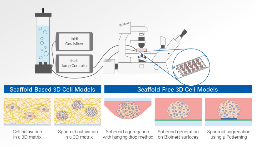
Each method provides different levels of control over cell aggregation, spatial organization, extracellular matrix composition, nutrient transport, imaging, and experimental throughput.
Cell Cultivation in a 3D Matrix
Hydrogel-based scaffolds provide a three-dimensional environment that recapitulates key properties of the extracellular matrix found in tissues. Within these scaffolds, single cells and multicellular aggregates can attach, migrate, proliferate, differentiate, and reorganize their surroundings. Commonly used matrices include collagen gels and basement membrane extracts such as Matrigel®.
Collagen is the most abundant protein in mammals and forms fibrous networks that reproduce important structural characteristics of the native extracellular matrix. Its concentration and polymerization conditions can be adjusted to influence matrix density, stiffness, and pore structure. Matrigel® is derived from Engelbreth-Holm-Swarm mouse sarcoma and contains extracellular matrix (ECM) proteins such as laminin and collagen IV, as well as a range of incompletely defined growth factors and other bioactive components. It polymerizes into a hydrogel at physiological temperatures, creating a 3D environment for cell culture and tissue engineering.
Both single cells and multicellular 3D aggregates, such as spheroids, organoids, and embryoid bodies, can be embedded and cultivated within a 3D matrix to investigate cell migration, morphology, cell–matrix interactions, differentiation, and tissue organization.
Application examples of matrix-based 3D cell culture are
- Organoid generation and expansion
- Tissue and tumor models
- Cell invasion assays
- Angiogenesis and tube formation
- Cancer–stroma co-culture
- Immune cell migration
- Tissue engineering
- Perfused MPS and organ-on-a-chip models
The use of ECM scaffolds is the current standard for the cultivation of organoids. Using hydrogels for 3D cell cultivation offers distinct advantages and disadvantages compared to scaffold-free methods like the hanging drop technique or the µ-Patterning technology. Hydrogels more closely replicate the extracellular matrix, providing a more physiological environment for cells, which is crucial for studying cell behavior, differentiation, and tissue development. Both, collagen and Matrigel®, can be modified in terms of stiffness and composition to suit different cell types and research needs. However, hydrogels have certain limitations. Unlike µ-Patterning, they do not allow for precise control over the spatial arrangement of cells, which can be crucial for certain applications like tissue engineering. Additionally, natural hydrogels may exhibit a batch-to-batch variability, potentially affecting the reproducibility of results.
3D cell aggregates often more accurately mirror in vivo conditions compared to single-cell cultures. However, it is also possible to culture and image single cells within a 3D gel, offering a unique perspective for various biological studies. This approach is particularly useful for investigating migration kinetics, cell-matrix-interactions and tube formation processes. In addition to cultures with only one cell type, the invasion behavior of two different cell types (e.g., cancer cells and fibroblasts) can be investigated by co-culturing them in the same vessel.
Although this method often does not match the complexity and fidelity of spheroid or organoid culture, it remains a cost-effective and straightforward alternative for research questions specifically centered on single-cell dynamics and responses.
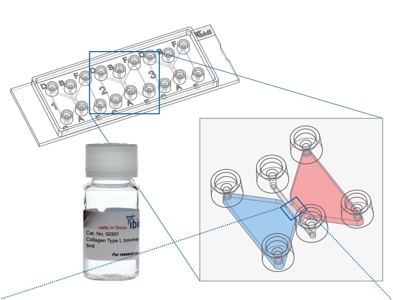
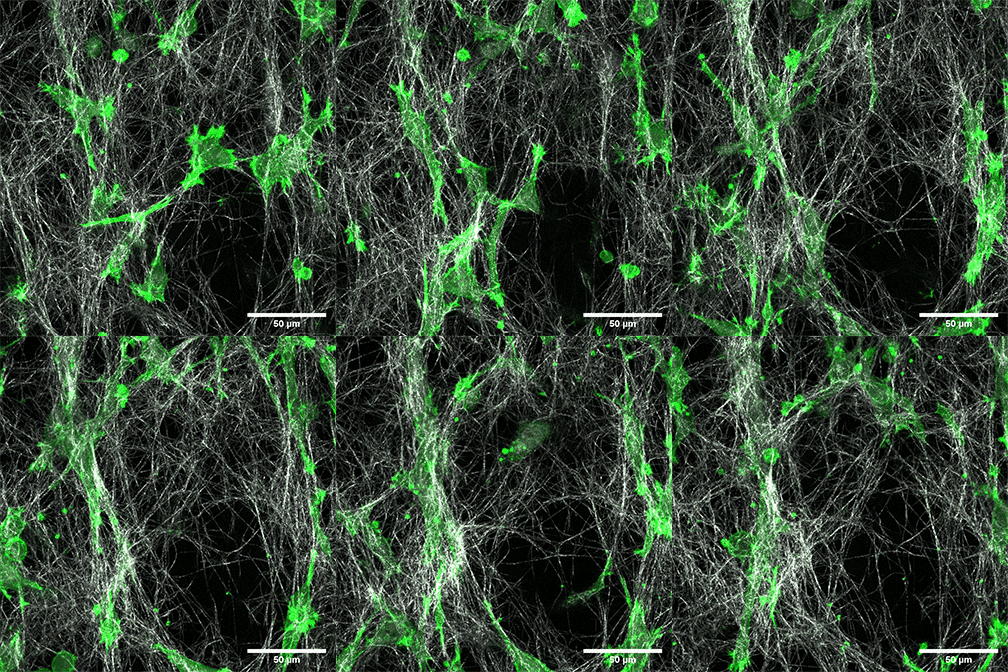
Individual LifeAct-expressing HT-1080 cells (green) in a Collagen Type I, Rat Tail layer in the µ-Slide Chemotaxis.
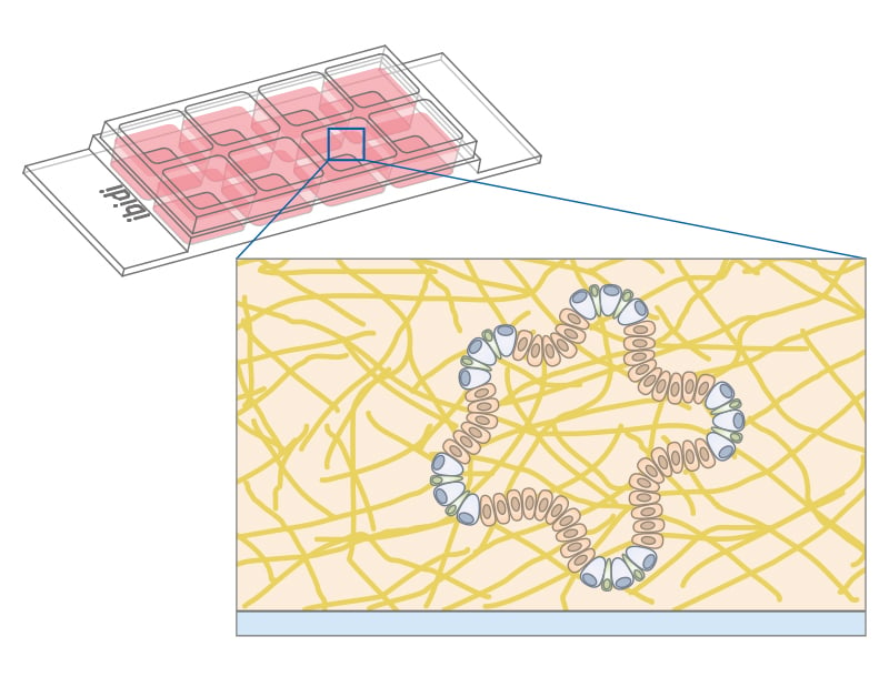
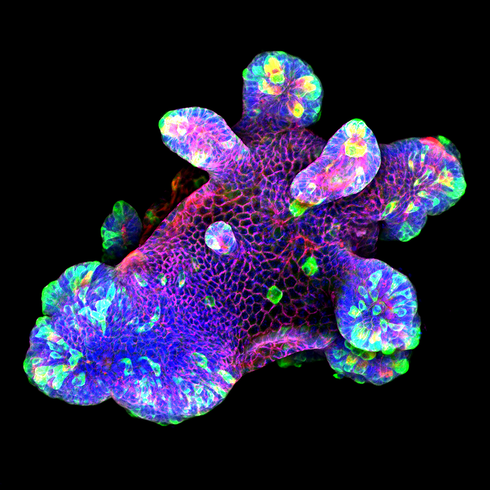
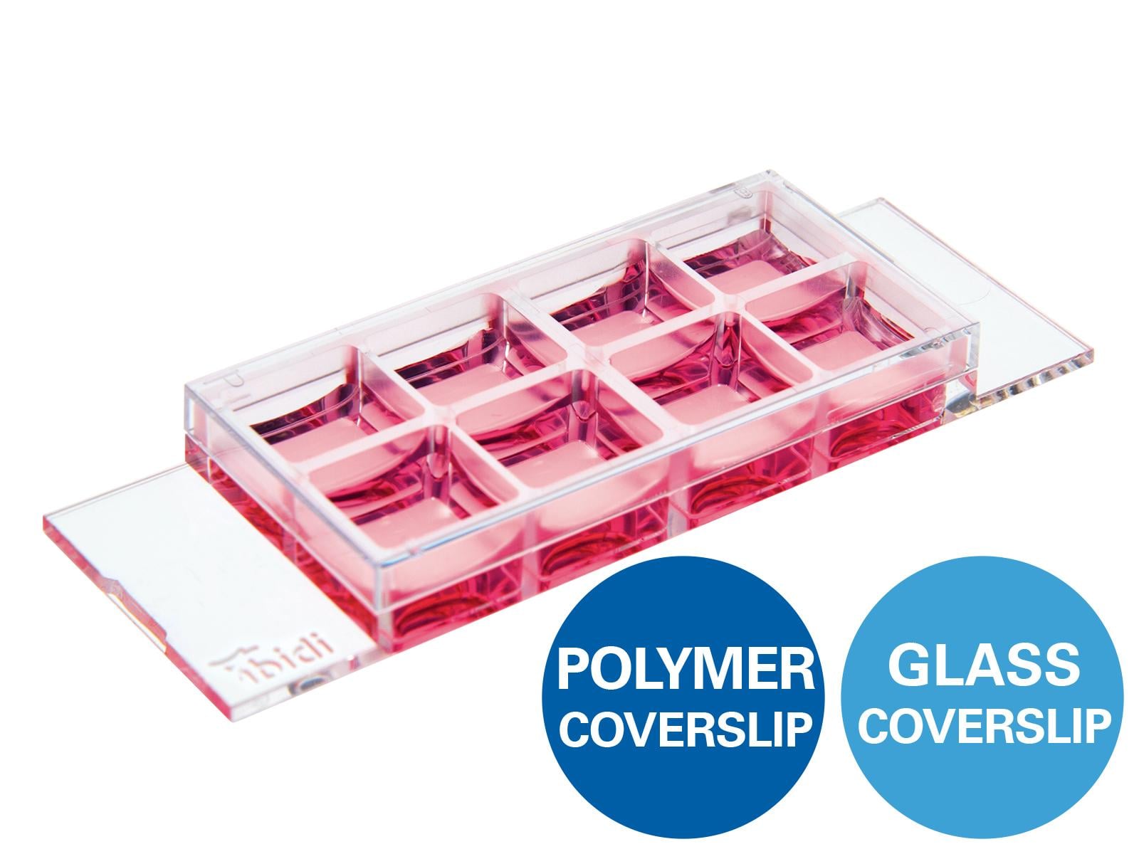
3D culture of an IL-22-treated mouse small intestine organoid grown in Matrigel® drops using an ibidi µ-Slide 8 Well. Image by Naveen Parmar, Norwegian University of Science and Technology (NTNU), Trondheim, Norway.
 | ibidi Blog Article |
Do you want to get an overview about the big variety of
3D Cell Culture Models? Then read the ibidi Blog.

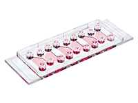
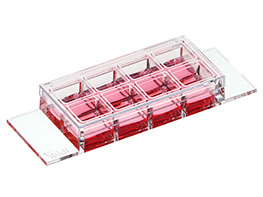
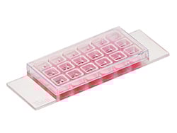
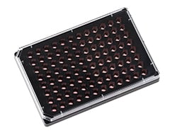
ibidi Solutions for Cell Cultivation in a 3D Matrix

Selected Publications for Cell Cultivation in a 3D Matrix
3D Chemotaxis assay with lymphoma cells in collagen matrix was done using the µ-Slide Chemotaxis.
Antonello P, Pizzagalli DU, Foglierini M, et al. ACKR3 promotes CXCL12/CXCR4-mediated cell-to-cell-induced lymphoma migration through LTB4 production. Front Immunol. 2023;13:1067885. doi:10.3389/fimmu.2022.1067885.
Read article
Murine mesenchymal-like colon cancer organoids were cultured in a 3D matrix and immunostained in the µ-Slide 15 Well 3D.
Poghosyan S, Frenkel N, Lentzas A, et al. Loss of Neuropilin-2 in Murine Mesenchymal-like Colon Cancer Organoids Causes Mesenchymal-to-Epithelial Transition and an Acquired Dependency on Insulin-Receptor Signaling and Autophagy. Cancers (Basel). 2022;14(3):671. doi:10.3390/cancers14030671.
Read article
An in vitro 3D model for cancer invasion was established in the µ-Slide 15 Well 3D.
Jouybar M, Sleeboom JJF, Vaezzadeh E, Sahlgren CM, den Toonder JMJ. An in vitro model of cancer invasion with heterogeneous ECM created with droplet microfluidics. Front Bioeng Biotechnol. 2023;11:1267021. doi:10.3389/fbioe.2023.1267021.
Read article
A liver-on-a-chip through hepatocyte 3D culture in a hydrogel was established using the µ-Slide III 3D Perfusion.
Christoffersson J, Aronsson C, Jury M, Selegård R, Aili D, Mandenius CF. Fabrication of modular hyaluronan-PEG hydrogels to support 3D cultures of hepatocytes in a perfused liver-on-a-chip device. Biofabrication. 2018;11(1):015013. doi:10.1088/1758-5090/aaf657.
Read article
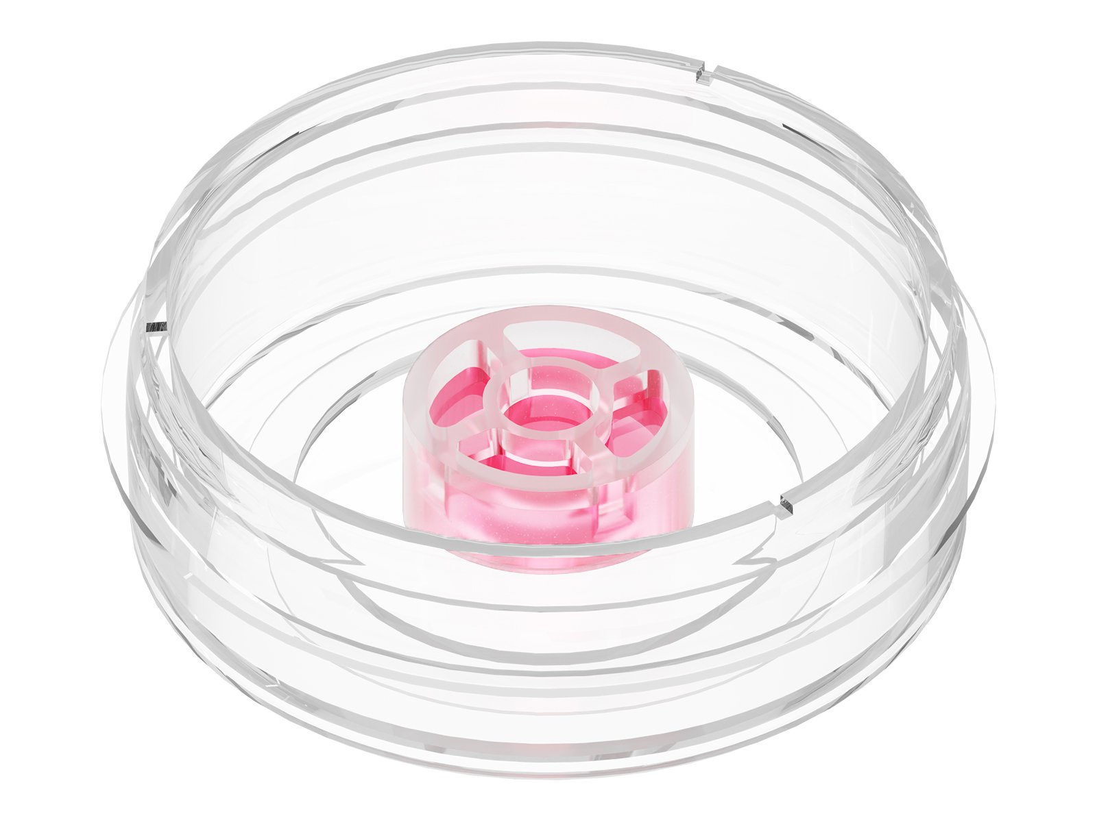
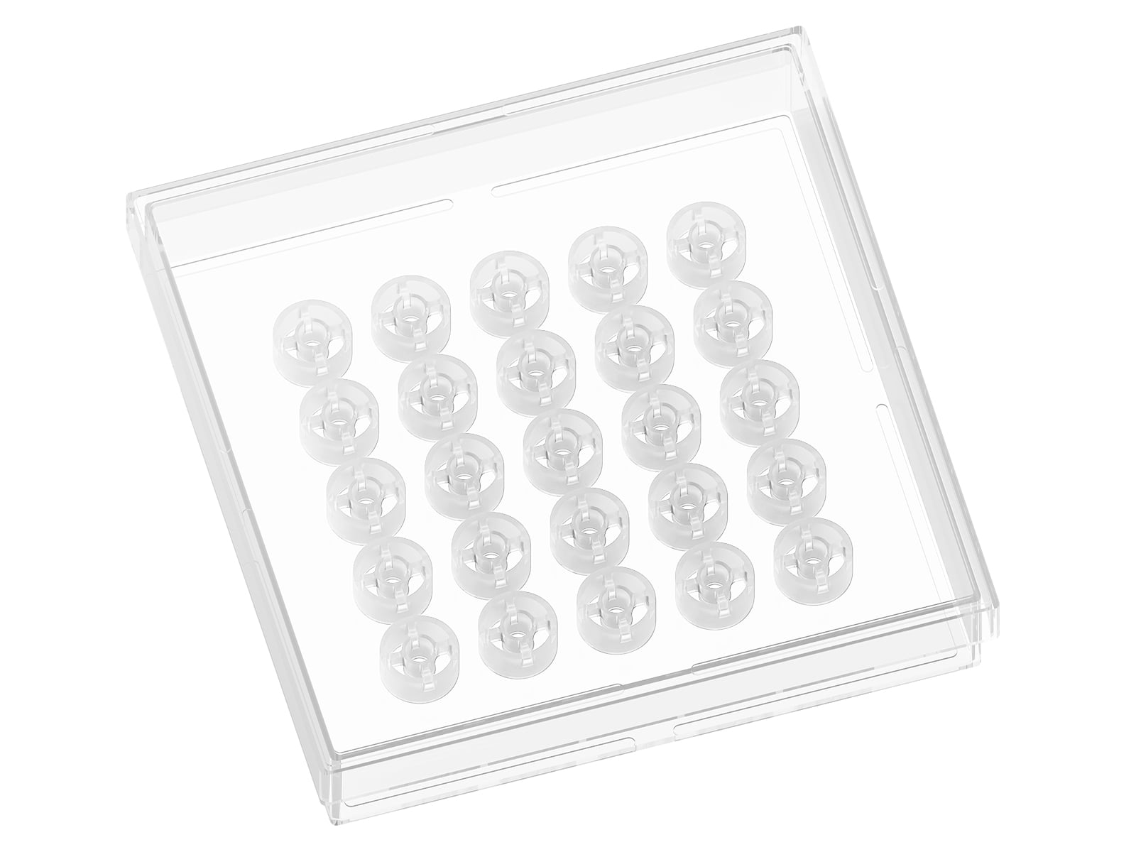
The hanging drop method, a traditional approach for the generation of uniform 3D aggregates, uses gravity to promote cell aggregation at the bottom of drops. This method involves placing small droplets of cell suspension on the underside of a culture dish lid. As gravity acts, cells within these droplets aggregate at the lowest point of the drop. This technique allows for precise control over the initial cell number in each droplet, leading to uniform spheroid size, an essential factor for consistent experimental results.
The hanging drop method is simple, cost-effective, and requires little specialized equipment or surface treatment. The small culture volume promotes efficient cell–cell interaction and can support the formation of compact, symmetrical aggregates. However, the restricted media volume makes media exchange and long-term cultivation more challenging. Handling individual droplets is labor-intensive, limits scalability, and increases the risk of disturbing or losing aggregates. In addition, aggregates often need to be transferred to another culture vessel for further cultivation, treatment, or high-resolution microscopy.
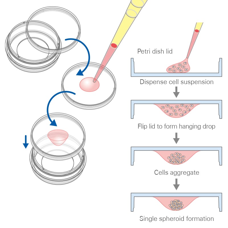
Principle of the hanging drop method. A defined volume of cell suspension with a known cell concentration is pipetted onto the inner surface of a cell culture dish lid. The lid is then inverted and placed onto the dish, allowing gravity to concentrate the cells at the lowest point of the hanging drop, where they aggregate into a compact 3D structure.
ibidi Solution for the Hanging Drop Method
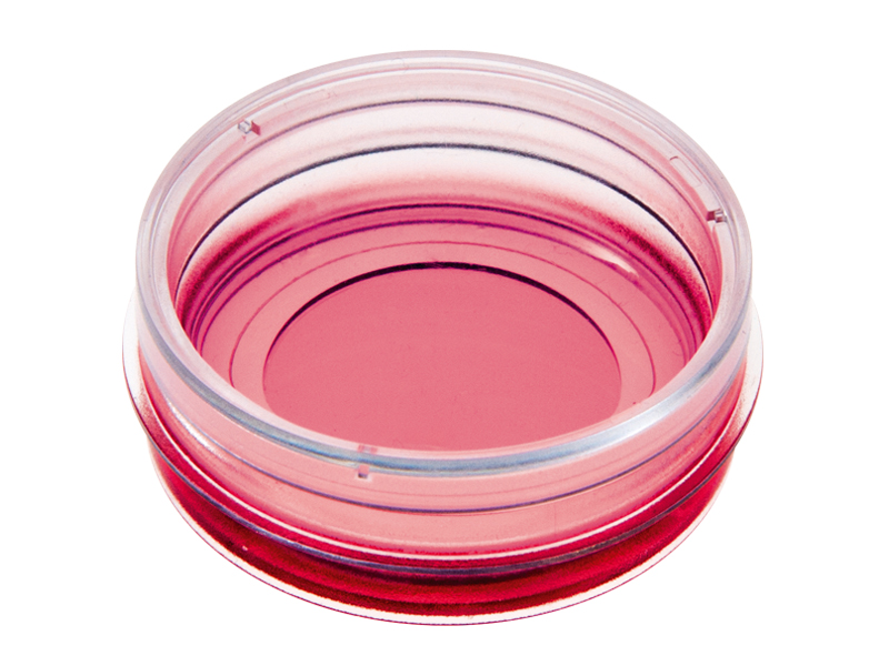
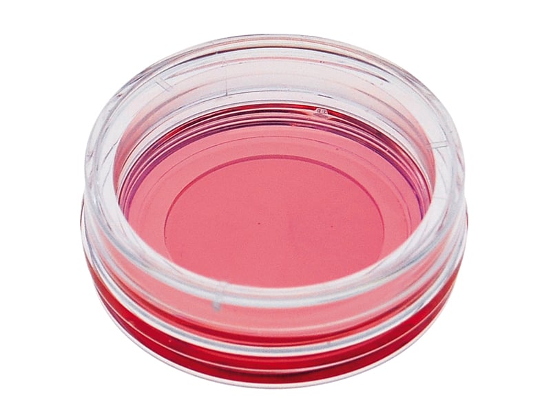
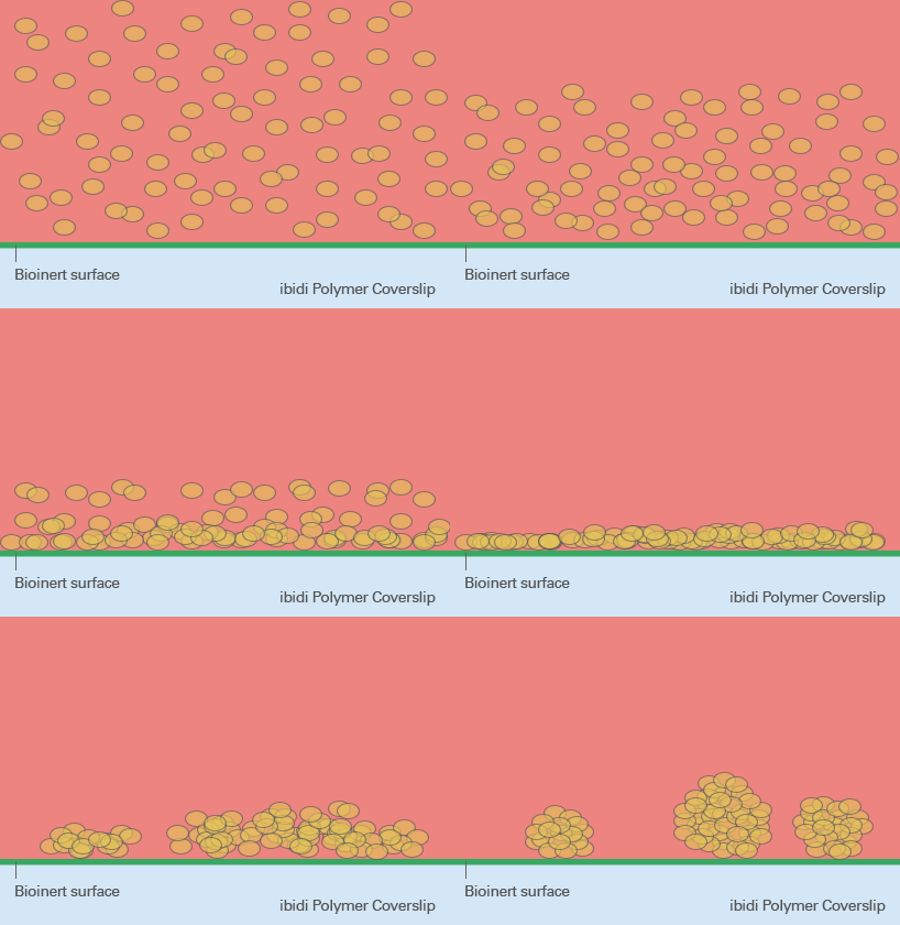
Bioinert and ultra-low attachment surfaces prevent cells from adhering to the culture vessel. Instead, suspended cells interact with one another and form three-dimensional aggregates. Depending on the cell source and culture protocol, this method can support the formation of spheroids, embryoid bodies, stem cell aggregates, and early-stage organoid structures.
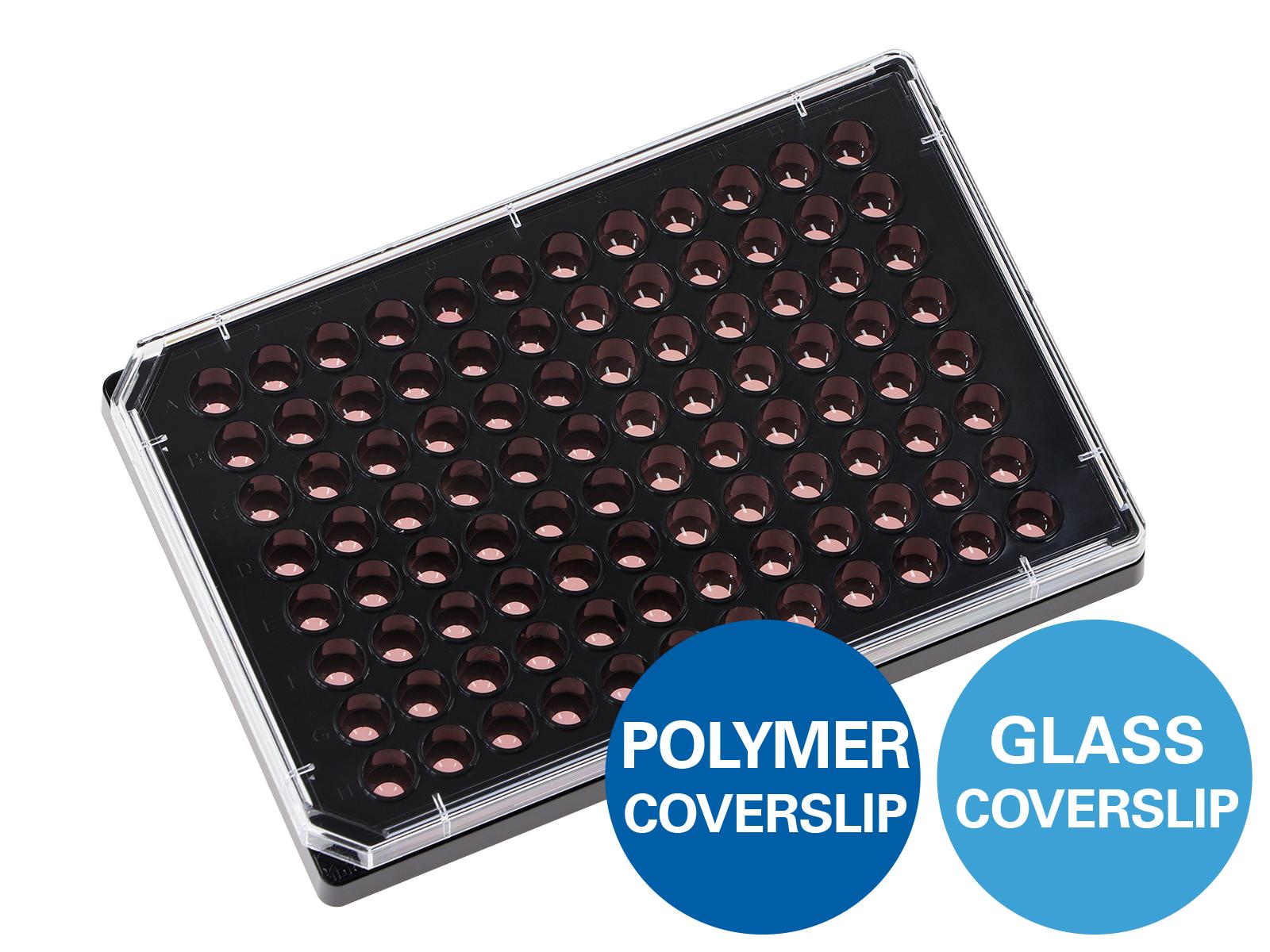
Bioinert surfaces are particularly useful when direct cell attachment to the surface would interfere with aggregation or differentiation. They provide a simple scaffold-free workflow, are compatible with many cell types, facilitate more natural cell-cell interactions, and allow flexible cultivation of 3D aggregates. Their integration into multiwell formats also facilitates parallel experiments, media exchange, treatment, downstream staining, and microscopy.
However, the number, size, shape, and position of the resulting aggregates may vary. These characteristics depend on factors such as the cell type, seeding density, vessel geometry, culture conditions, and strength of cell–cell adhesion.
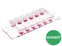
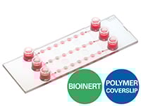
ibidi Solutions for 3D Cell Aggregation on Bioinert Surfaces

Selected Publications for 3D Cell Aggregation on Bioinert Surfaces
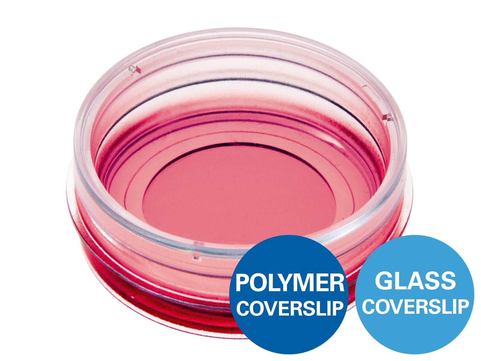
Hepatocellular carcinoma spheroids were generated and stained in the µ-Dish 35 mm, high Bioinert.
Bergamini C, Leoni I, Rizzardi N, et al. MiR-494 induces metabolic changes through G6pc targeting and modulates sorafenib response in hepatocellular carcinoma. J Exp Clin Cancer Res. 2023;42(1):145. doi:10.1186/s13046-023-02718-w.
Read article
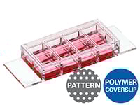
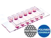
The ibidi µ-Patterning technology provides spatially defined ibiTreat adhesive patterns for confined cell arrays on a non-adhesive Bioinert surface. Cells attach only within the specified patterns, allowing controlled positioning and organization before or during aggregate formation.
This approach can be used to generate spatially defined spheroids and other multicellular aggregates. Depending on the cell type and differentiation protocol, it may also support embryoid body formation or provide defined starting structures for more complex 3D models. Because the aggregates remain at defined positions, the same structures can be repeatedly located during phase contrast, fluorescence, or time-lapse microscopy. This makes micropatterning particularly valuable for longitudinal analysis, automated imaging, and screening workflows.
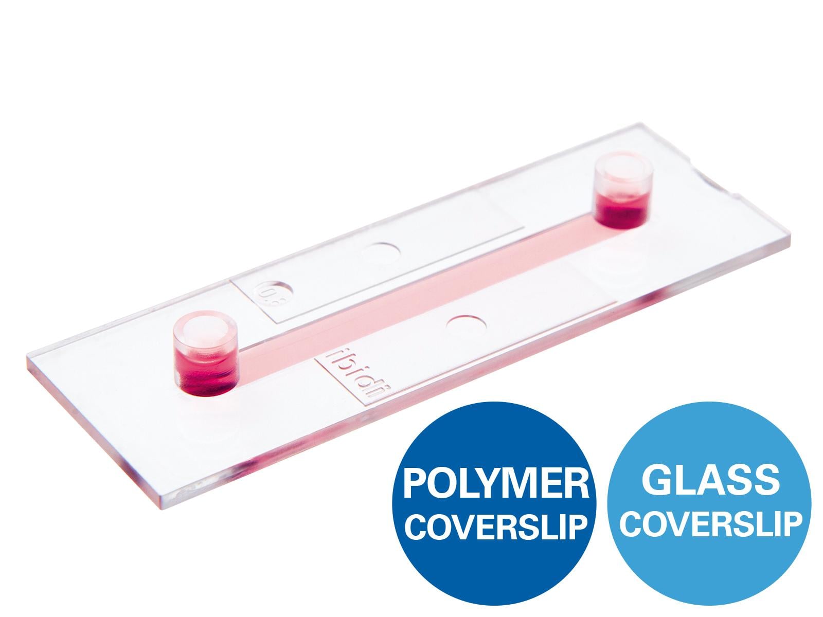
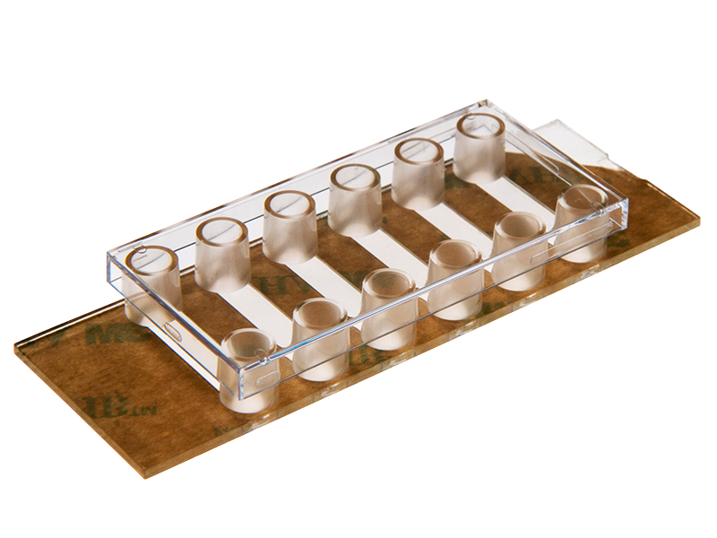
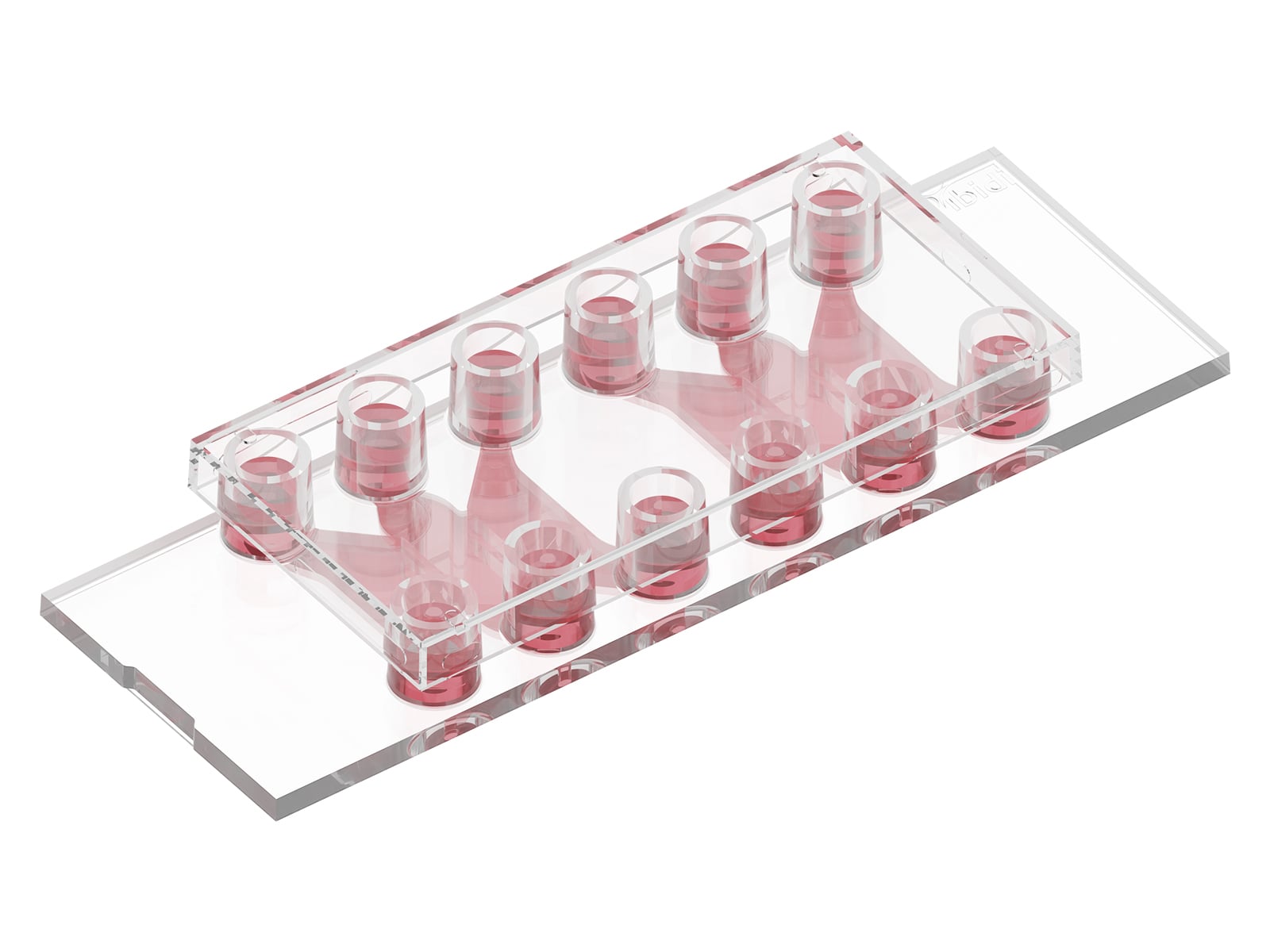
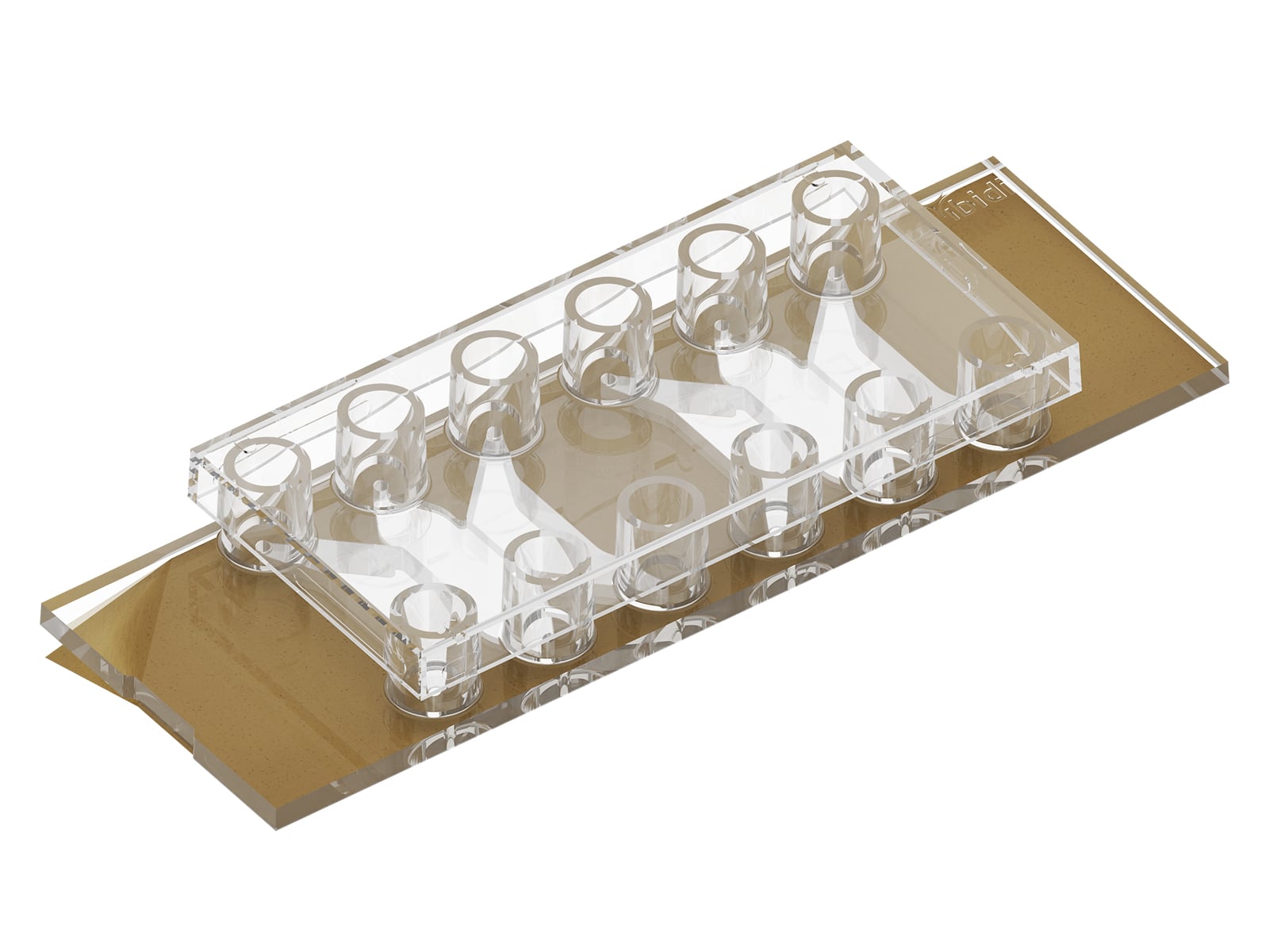
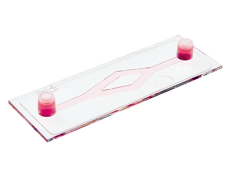
Combining the µ-Patterning technology with the µ-Slide 8 Well high and µ-Slide VI 0.4 enables aggregate generation, cultivation, treatment, staining, and imaging in the same vessel, reducing transfer steps and minimizing the risk of losing or damaging the samples.
However, aggregate formation and morphology still depend on factors such as the selected cell type, seeding density, cell–cell adhesion, pattern dimensions, and culture conditions. Not all cells form compact or homogeneous aggregates equally well, and the predefined pattern geometry may need to be optimized for each model. In addition, µ-Patterning is best suited for applications that benefit from controlled adhesion and spatial positioning. It may be less appropriate when freely suspended aggregates or complete detachment from the culture surface are required.
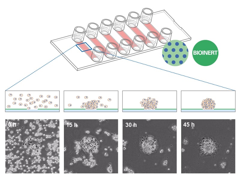
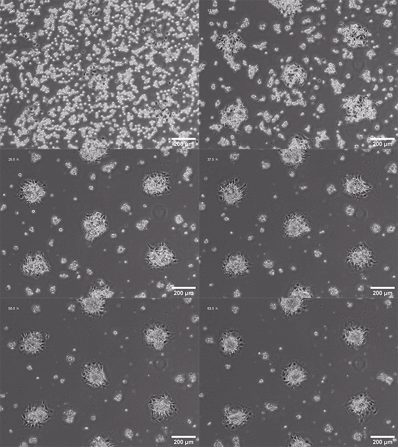
Spheroid formation of NIH-3T3 cells (murine embryo fibroblasts) on 200 µm adhesion spots using ibidi µ-Patterning. Spheroid generation was documented for 64 hours. Phase contrast live cell imaging, 4x objective lens.
ibidi Solutions for 3D Cell Aggregation Using µ-Patterning
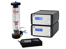
Spheroids, organoids, embryoid bodies, and engineered tissues can be integrated into microphysiological systems (MPS) and organ-on-a-chip models. These platforms recreate more physiologically relevant conditions by providing controlled media flow, nutrient and oxygen supply, mechanical stimulation, or defined tissue interfaces. Such dynamic culture systems enable the investigation of barrier function, tissue interactions, drug responses, and other physiological processes under conditions that more closely resemble the in vivo environment.
Compared with static culture, MPS and organ-on-a-chip models provide greater physiological relevance and allow precise control over the cellular microenvironment. However, they typically require more complex experimental setups, specialized equipment, and careful optimization of flow conditions and culture protocols.
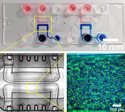
PTubeChip – 3D-printed organ models with perfused microchannels
Learn more about advanced 3D cell culture methods, including perfusion, bioprinting, and organ-on-a-chip models here.
ibidi Solutions for Organ-on-a-Chip Applications


Read on and have a look at methods for Advanced Cultivation of 3D Cell Models or Experimental Applications.