


Live Cell Imaging: Investigate Cell Biology in Real Time
Live cell imaging is the time-lapse microscopy of dynamic processes in living cells. It enables observation of cell-cell interactions, the behavior of single cells, and the dynamics of cell organelles or cellular molecules. Several imaging techniques can be applied, such as phase contrast microscopy, fluorescence and confocal microscopy, multiphoton microscopy, light sheet microscopy, or even TIRF and super-resolution microscopy.
Many topics in cell biology can be addressed using live cell imaging:
- Investigating cell migration in chemotaxis assays or wound healing assays, for example
- Mimicking blood or lymphatic vessels using cell culture under flow
- Studying angiogenesis with tube formation assays
- Measuring cell proliferation over time
- Analyzing inter- and intracellular signaling using specific fluorescence staining and high-resolution microscopy
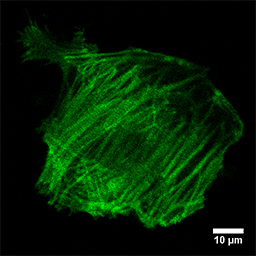
- Gaining insight into the cytoskeletal dynamics
High-resolution live cell imaging for the visualization of the contraction rates of cardiomyocytes, plated in a µ-Dish 35 mm, high ibiTreat (coated with Fibronectin). F-Actin is stained with LifeAct. 100x objective lens.
During the whole live cell imaging experiment, the cells need to be kept alive and healthy. Therefore, physiological conditions have to be established and maintained on the microscope. For both the researcher and the equipment, this presents several challenges and requires exact experimental planning. However, live cell microscopy offers many novel possibilities for achieving a better understanding of the biological dynamics within the cell.
Read on and learn more about the Parameters for Live Cell Imaging or the requirements for a Live Cell Imaging Experiment.
ibidi Blog Article |
Manuel Izquierdo from the Universidad Autónoma de Madrid communicates live cell imaging approaches to get "Mechanistic Insights into Immune Synapse Formation During Immune Response"
See cancer dynamics unfold through live imaging in "From Tumor Microenvironment to Microscope: Capturing Cancer in Motion"