Since many cellular processes can be visualized directly on the microscope, different imaging techniques are required.
Some of these experiments, such as rolling adhesion, chemotaxis, or tube formation assays, do not require any staining and can be easily imaged using light microscopy (e.g., phase contrast). Even though light microscopy illumination has only a little phototoxicity, it should still be minimized. For example, the light should only be switched on during image acquisition. Additionally, the necessary light intensity can be minimized by using sensitive cameras.
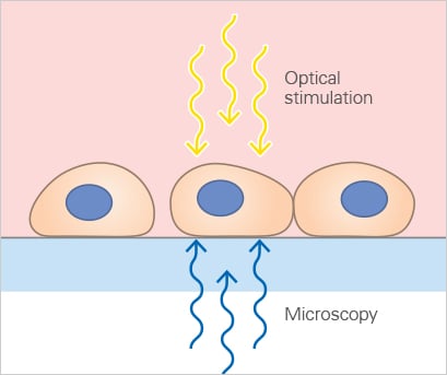
However, fluorescence microscopy that uses reporter proteins like GFP will reveal more structural details and molecular interaction. When using fluorescence-based microscopy techniques (e.g., confocal microscopy), it is especially crucial to minimize the cell stress that is caused by excitation light. Phototoxic effects and photobleaching happen quickly in living cells and might alter the outcome of the experiment.
Find more details about the different live cell microscopy requirements and techniques, fluorescence staining techniques and how to prevent photobleaching.