


Phase Contrast Microscopy:
Principle, Advantages, and Applications
Phase contrast microscopy is a widely used optical imaging technique to visualize transparent, unstained cells. In cell biology, it enables high-contrast imaging of cell morphology, dynamics, and behavior in real time. It is a standard method in live cell imaging workflows where minimal sample preparation and non-invasive observation are required.
What Is Phase Contrast Microscopy?
Phase contrast microscopy is a label-free imaging technique that enhances contrast in transparent specimens by transforming phase shifts in light into intensity differences. These phase shifts occur when light passes through materials with different refractive indices, such as cellular structures. Instead of requiring dyes or fluorescent markers, the method uses specialized optical components to make otherwise invisible structures clearly visible. This makes it ideal for imaging living cells in their natural state, preserving biological function and reducing experimental artifacts.
When to Use Phase Contrast Microscopy
Phase contrast microscopy is the preferred choice when a fast, simple, and non-disruptive imaging method is needed, especially for routine live cell analysis.
Use it when:
- Imaging living, unstained cells in real time
- Studying cell morphology, growth, and dynamics
- Performing long-term time-lapse experiments with low phototoxicity
- Label-free observation is required
- Fluorescence labeling is unnecessary or should be avoided
For higher contrast imaging of fine structures, DIC microscopy may be more suitable. If fluorescence labeling or multi-channel imaging is required, widefield microscopy is typically the better choice.
How Does Phase Contrast Microscopy Work?
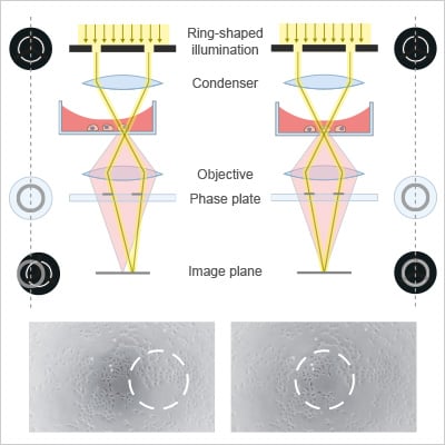
Unstained living cells absorb practically no light and therefore appear nearly transparent under conventional brightfield microscopes. However, as light passes through these cells, it experiences small phase shifts due to differences in refractive indices between cellular components and the surrounding medium, which are almost invisible to the human eye. A phase contrast microscope transforms these small phase shifts into variations in light intensity, resulting in high-contrast images.
This is achieved using both an annular ring located in the condenser and a phase ring located in the back focal plane of the objective. The annular ring creates a tube-shaped illumination, which is selectively shifted by the phase ring in the objective (usually by ±90° (λ/4)) to interfere constructively or destructively with scattered light, coming from the sample. The resulting amplitude changes reveal otherwise invisible cellular structures.
Comparison: Phase Contrast vs Brightfield Microscopy
Brightfield and phase contrast microscopy are widely used light microscopy techniques for imaging cells, each optimized for different contrast needs and experimental conditions.
When to use phase contrast:
- Imaging living, unstained cells
- Observing cell morphology and internal structures
- Performing time-lapse live cell imaging
- When label-free imaging is required
When to use brightfield:
- When colorimetric stains (e.g., H&E) are used
- High-contrast samples that are naturally visible
| Feature | Phase Contrast | Brightfield |
| Contrast | High (unstained cells) | Low without staining |
| Sample prep | No preparation needed | Often requires staining |
| Live imaging | Excellent | Limited |
| Cell visibility | Clear internal structures | Poor for transparent cells |
| Phototoxicity | Low | Low |
What Are the Main Applications of Phase Contrast Microscopy?
Phase contrast microscopy is widely used for live cell imaging and monitoring, enabling detailed analysis of cell morphology, migration, and proliferation. It is particularly valuable in time-lapse experiments, as well as in cancer and stem cell research to study cell behavior and dynamics in a label-free, non-invasive manner.
Advantages and Limitations of Phase Contrast Microscopy
Phase contrast microscopy is a widely used, cost-effective, and non-destructive imaging technique that enables label-free observation of living cells. It requires no staining or labeling and therefore minimizes sample preparation. Additionally, phase contrast minimizes phototoxicity due to low light intensity, making it ideal for long-term, real-time imaging.
However, it can produce halo artifacts, offers limited molecular specificity, and provides lower contrast compared to fluorescence microscopy. Additionally, it is less suitable for imaging thick samples.
How Can I Optimize My Phase Contrast Images?
The quality of phase contrast images depends strongly on the precise alignment of the annular ring with the phase ring. However, even with perfect alignment and the elimination of other mechanical disturbances, optical aberrations (such as those caused by meniscus formation) can degrade image quality. The meniscus of the cell culture medium at the air–liquid interface acts as a lens, disrupting the precisely aligned light path of the microscope. This effect is particularly pronounced in small wells, such as those in standard 96-well plates, where the meniscus curvature is more pronounced. In summary, achieving high-contrast images requires (i) precise alignment of the phase and annular rings to optimally adjust the light path and (ii) the use of suitable labware to minimize interference from the meniscus effect.
Beam path with meniscus | Beam path without meniscus |
ibidi Solutions for Enhanced Phase Contrast Imaging
High-quality phase contrast imaging depends on minimizing optical artifacts, especially the meniscus effect, which causes uneven illumination and poor contrast. Using optimized labware ensures a flat imaging surface, consistent optical conditions, and reproducible results. ibidi provides specialized solutions designed to eliminate meniscus-related distortions and improve overall image quality. Available with either #1.5 Polymer Coverslip Bottom or 1.5H Glass Coverslip Bottom, both deliver exceptional optical quality and high-contrast phase contrast imaging.
ibidi provides specialized solutions designed to eliminate meniscus-related distortions and improve overall image quality. Available with either #1.5 Polymer Coverslip Bottom or 1.5H Glass Coverslip Bottom, both deliver exceptional optical quality and high-contrast phase contrast imaging.
ibidi Channel µ-Slides
The ibidi Channel μ-Slides provide optimal conditions for phase contrast microscopy by completely preventing meniscus formation.
- No air-water interface; no meniscus
- Uniform contrast across the entire channel
- Compatible with the ibidi Pump System for flow applications
Key benefits: Consistent, artifact-free imaging across the full field of view, with integrated perfusion capability for dynamic live cell experiments.
For detailed information, please refer to "Phase Contrast in Channel Slides" or read our Application Note 03 (PDF).
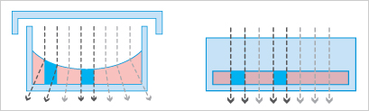
Standard well Strong meniscus at the air-liquid interface; poor phase contrast especially near the walls | ibidi Channel Slides No meniscus for good phase contrast over the whole observation area |
ibidi µ-Slides Ph+
The ibidi µ-Slides Ph+ are designed to reduce meniscus effects in standard well formats.
- Integrated design minimizes liquid curvature
- Uniform phase contrast across the entire well
- Improved reproducibility compared to standard plates
Key benefits: Reliable imaging across the whole well, not just the center.
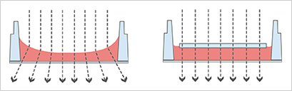
Standard well Strong meniscus at the air-liquid interface; poor phase contrast especially near the walls | µ-Slides Ph+ No meniscus for good phase contrast over the whole observation area |
ibidi µ-Slide 15 Well 3D and µ-Plate 96 Well 3D
The µ-Slide 15 Well 3D and µ-Plate 96 Well 3Dare optimized for 3D and hydrogel-based cultures, where meniscus effects are even more pronounced.
- “Well-in-a-well” design prevents gel meniscus formation
- Cells remain in a single focal plane
- Suitable for high-throughput and 3D assays
Key benefits: Clear phase contrast imaging even in complex 3D environments.
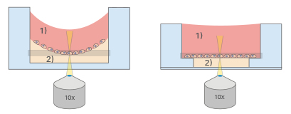
Standard well Meniscus at air-liquid interface (1) and meniscus formation of the gel surface (2); poor phase contrast and not possible to focus on all cells simultaneously. | µ-Slide 15 Well 3D / µ-Plate 96 Well 3D Planar air-liquid interface (1) and meniscus-free gel surface (2) for good phase contrast with all cells on one focal plane |
Common Problems and Troubleshooting
Why is my phase contrast image low in contrast?
Low contrast is often caused by misalignment of the condenser annulus and phase ring or incorrect phase settings.
Solution: Realign the condenser annulus carefully and ensure the correct phase ring is selected. Proper alignment is critical for optimal contrast.
Find more information how to align your phase contrast microscope here.
Why is my illumination uneven?
An inhomogeneous illumination can be caused by the meniscus effect, when the liquid surface in a well or chamber is curved. This leads to uneven illumination and image artifacts, especially toward the edges.
Solution: Whenever possible, use large vessels and image near the center of the well (avoid edges where the meniscus is strongest). Or use specialized vessels such as the ibidi phase contrast labware.
Why is my image blurry or not sharp?
Blurry images are typically caused by incorrect focus, uneven sample thickness, or suboptimal imaging surfaces.
Solution: Refocus carefully, use vessels with optimal optical thickness, and ensure cells are evenly distributed.
Why do I see halos around my cells?
Halo artifacts are inherent to phase contrast microscopy and occur due to the way light interference is generated at edges of structures.
Solution: Slightly adjust focus and illumination to minimize halos. If halos interfere with analysis, consider alternative imaging methods such as DIC.
FAQs
What is phase contrast microscopy used for?
It is used to visualize live, unstained cells and analyze cell morphology, movement, and behavior in real time.
Why use phase contrast instead of staining?
It allows non-invasive imaging of living cells without altering or damaging the sample.
What is the principle of phase contrast microscopy?
It converts phase shifts in light passing through a sample into visible intensity differences.
Can phase contrast be used for time-lapse imaging?
Yes, it is widely used for long-term live cell imaging due to low phototoxicity.
Can phase contrast microscopy provide quantitative or 3D information?
Standard phase contrast microscopy provides qualitative contrast only. For quantitative or 3D-like data, advanced methods such as quantitative phase imaging (QPI) measure phase shifts to generate maps of optical thickness and refractive index in a label-free manner.
How does quantitative phase imaging (QPI) work?
QPI measures phase shifts by interfering light from the sample with a reference beam, enabling quantitative maps of optical thickness and refractive index.
References
F. Zernike. "Phase contrast, a new method for the microscopic observation of transparent objects". Physica, 1942, part I: 10.1016/S0031-8914(42)80035-X, part II: 10.1016/S0031-8914(42)80079-8.
read abstract part I / part II
E. Horn, R Zantl. Phase-Contrast Light Microscopy of Living Cells Cultured in Small Volumes. Microsc Anal, 2006, 20(3):5–7
read abstract

Article written by Stefanie Kiderlen, PhD
ibidi GmbH | September 5, 2025
Cell biologist and microscopy specialist with expertise in advanced imaging, live cell imaging, and 3D cell culture models. Stefanie received her PhD at the Biophysical Department at LMU, Munich, focusing on atomic force microscopy in 2D and 3D cell culture systems.