



FRAP Microscopy:
Principle, Workflow, and Applications
Fluorescence Recovery After Photobleaching (FRAP) is a fluorescence microscopy technique used to estimate diffusion and mobility parameters in living cells. FRAP is especially useful for studying membrane proteins, cytoskeletal structures, intracellular transport, and biomolecular interactions in live cell imaging experiments. It is commonly performed with confocal microscopy and fluorescently labeled molecules such as GFP-tagged proteins.
What Is FRAP Microscopy?
FRAP is a fluorescence microscopy method used to analyze how fluorescently labeled molecules move within cells, membranes, tissues, or biomaterials. In a FRAP experiment, a defined region of interest (ROI) is photobleached with high-intensity light, and the return of fluorescence is monitored over time as unbleached molecules move into the bleached area. This can provide information about diffusion rates, mobile and immobile fractions, and molecular exchange dynamics.
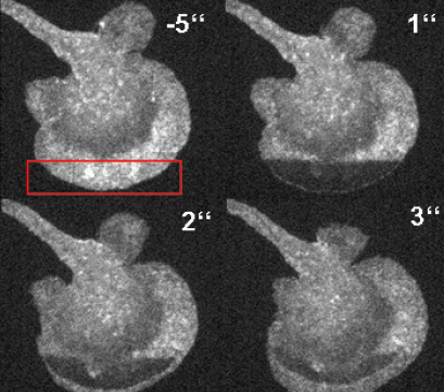
FRAP-based visualization of the F-actin diffusion in a primary dendritic cell.
How Does FRAP Work?
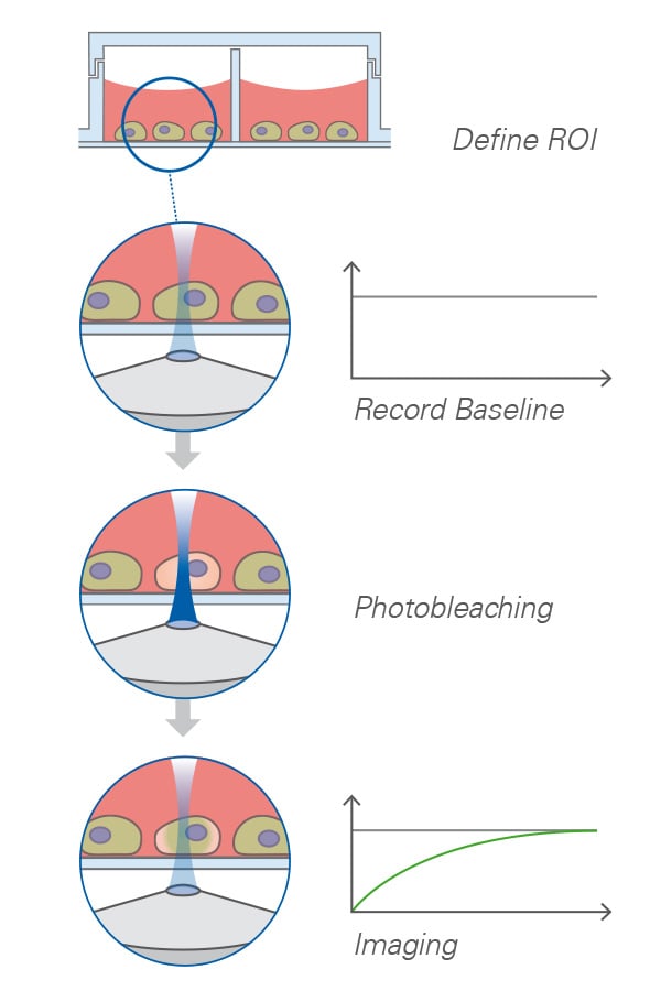
In FRAP microscopy, a fluorescent molecule of interest is first imaged at low light intensity to record its baseline fluorescence. A defined ROI is then exposed to a short exposure of high-intensity laser light. This photobleaches the fluorophores in that area and creates a dark region in the fluorescent sample. Over time, unbleached molecules from surrounding areas move into the bleached ROI, causing fluorescence to recover. The speed and extent of this recovery are plotted as a fluorescence recovery curve. This curve can be used to estimate parameters such as diffusion coefficient, recovery half-time, and mobile fractions.
When to Use FRAP Microscopy
Use FRAP when the main goal is to understand how fast and how freely molecules move in living cells or biological samples. FRAP is especially useful:
- When studying protein mobility and dynamics in live cells
- When measuring diffusion in membranes, cytoplasm, nucleus, organelles, or biofilms
- When comparing mobile and immobile molecular fractions
- When investigating binding, unbinding, trafficking, or molecular exchange
- When testing how drugs, mutations, or environmental conditions affect molecular dynamics
FRAP is less suitable when the sample is highly light-sensitive, when fluorescent labeling alters molecular behavior, or when recovery is too fast or too slow for the imaging setup.g.
Typical FRAP Workflow
Step 1: Prepare fluorescently labeled cells or samples
Use a fluorescent protein, dye, or probe that specifically labels the molecule or structure of interest.
Step 2: Select the imaging region
Choose a suitable cell, membrane area, organelle, or subcellular structure and define the FRAP region of interest.
Step 3: Record pre-bleach images
Acquire baseline fluorescence images at low illumination intensity to avoid unwanted photobleaching.
Step 4: Photobleach the ROI
Apply a short, high-intensity laser pulse to bleach fluorescence in the selected region.
Step 5: Monitor fluorescence recovery
Record a time series after bleaching to measure how fluorescence returns to the ROI.
Step 6: Normalize and analyze the recovery curve
Correct for background, acquisition bleaching, and initial intensity, then calculate recovery half-time, mobile fraction, immobile fraction, and diffusion-related parameters.
Step 7: Compare biological conditions
Use FRAP curves to compare treatments, mutations, cell types, membrane domains, or environmental conditions.
Which Microscope Is Used for FRAP?
FRAP is commonly performed with confocal fluorescence microscopy, because confocal systems allow precise laser bleaching of a defined region of interest and reduce out-of-focus fluorescence during recovery imaging. This is especially useful for FRAP experiments in thicker samples, intracellular structures, or 3D biological environments. However, FRAP is not limited to confocal microscopy. It can also be performed using widefield fluorescence microscopes, provided that controlled photobleaching and time-lapse imaging are possible. However, compared to confocal systems, widefield FRAP offers less precise spatial control of the bleached region and may be affected by out-of-focus fluorescence, which can influence the accuracy of quantitative analysis.
What Are the Advantages and Limitations of FRAP?
Advantages of FRAP:
- Enables live cell analysis of molecular mobility
- Provides quantitative information from fluorescence recovery curves
- Suitable for membrane, cytoplasmic, nuclear, and organelle studies
- Compatible with fluorescent proteins such as GFP-tagged constructs
- Can reveal mobile and immobile molecular fractions
Limitations of FRAP:
- Requires fluorescent labeling of the molecule of interest
- Photobleaching can damage sensitive live cells
- Very fast recovery may be difficult to capture
- Very slow recovery can require long-term live cell stability
What Are the Applications of FRAP?
Protein Mobility in Live Cells
FRAP is widely used to study how proteins move within the cytoplasm, nucleus, membranes, or organelles. It helps determine whether proteins are freely diffusing, transiently bound, or part of larger molecular complexes.
Membrane Protein Diffusion
FRAP is commonly applied to analyze lateral diffusion in cell membranes and artificial membrane systems. This is useful for studying receptor mobility, membrane organization, and lipid–protein interactions.
Cytoskeletal Dynamics
FRAP can be used to investigate dynamic structures such as actin, microtubules, and associated proteins. Recovery behavior can reveal turnover rates and structural stability.
Intracellular Transport
FRAP helps analyze molecular exchange between cellular compartments, including the nucleus, cytoplasm, vesicles, and organelles.
Drug Discovery and Treatment Response
FRAP can show how drugs, inhibitors, or mutations affect protein mobility, binding behavior, and cellular transport mechanisms.
ibidi Solutions for FRAP Microscopy
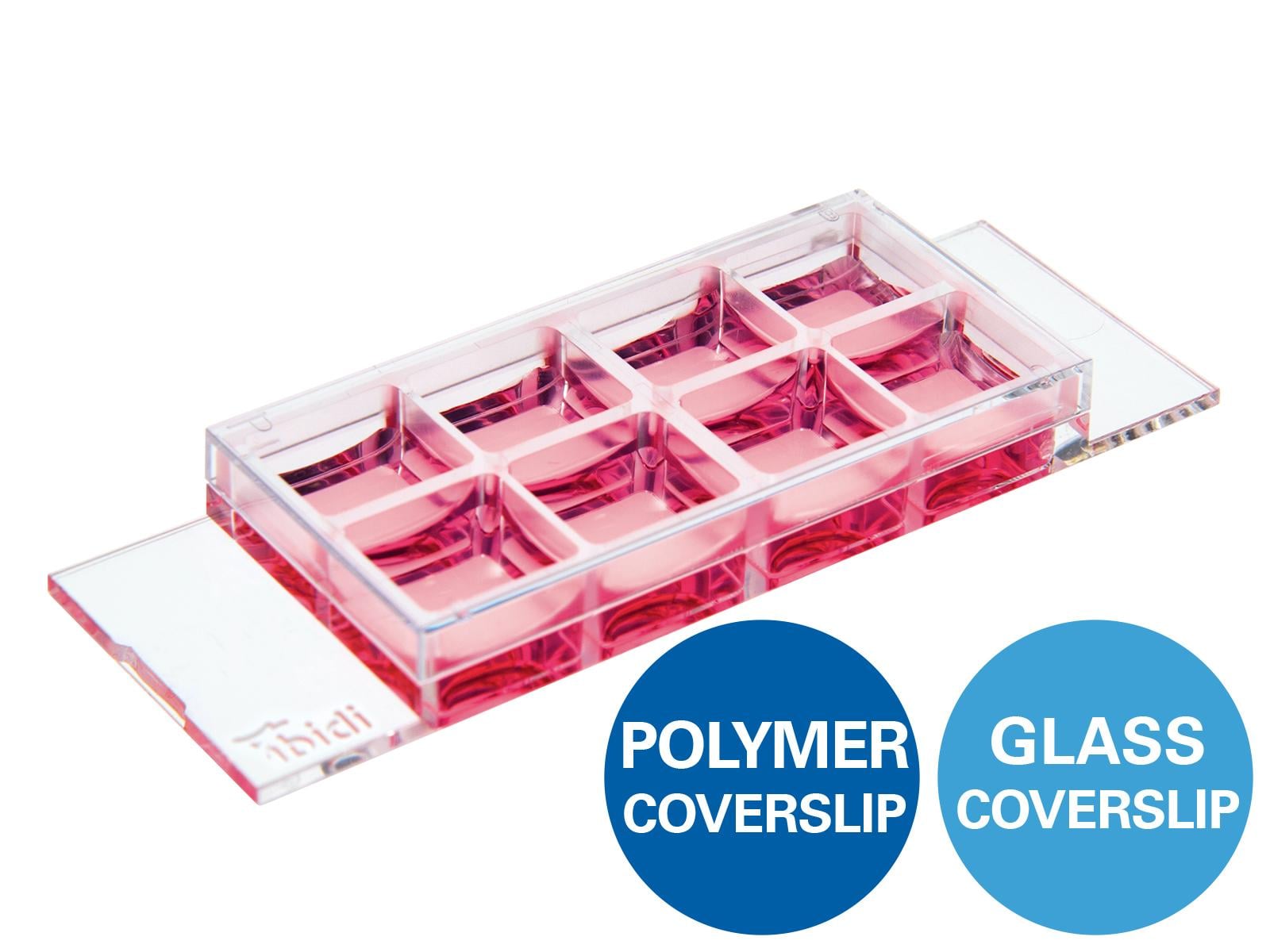
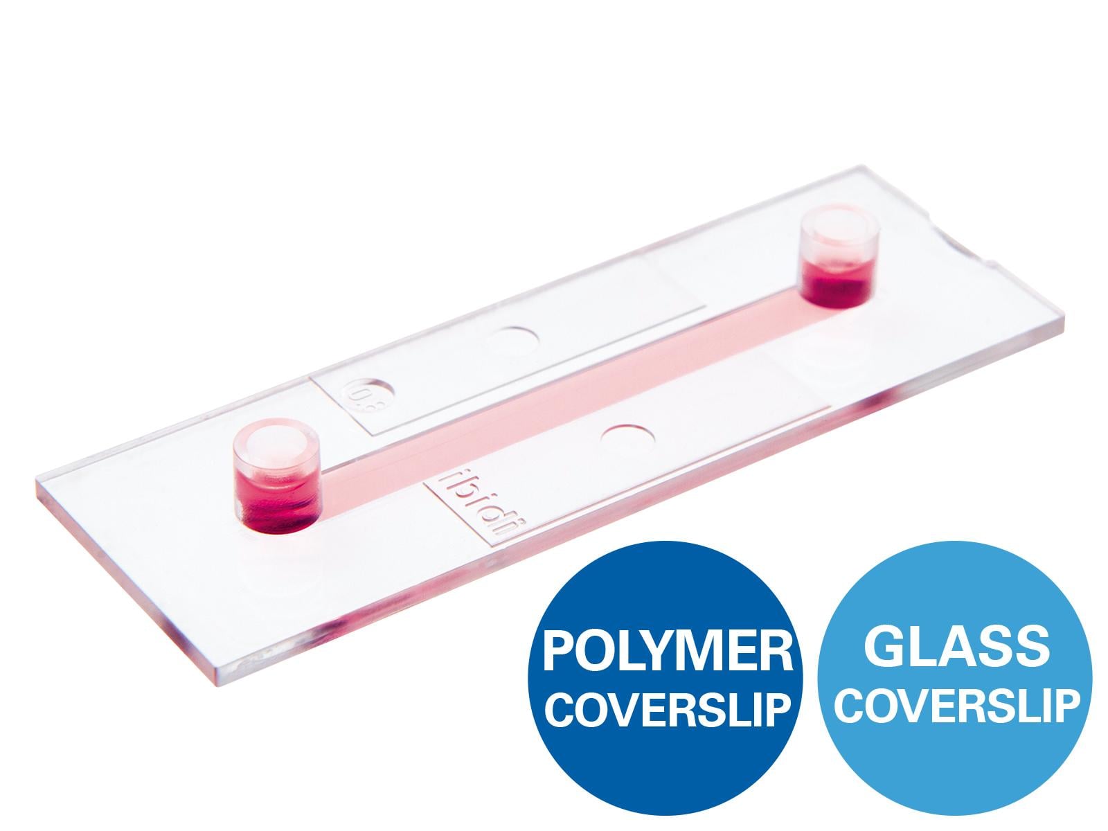
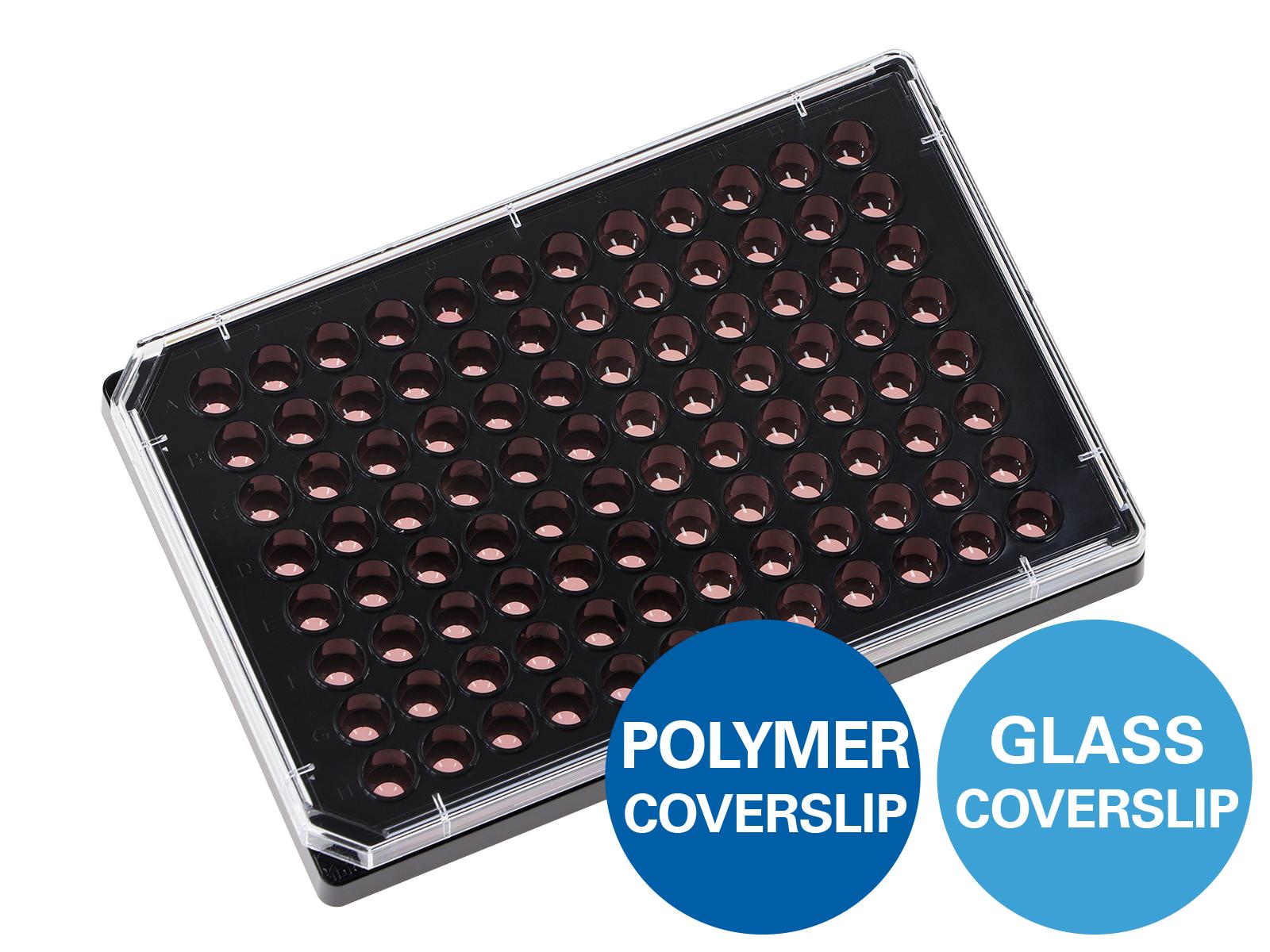
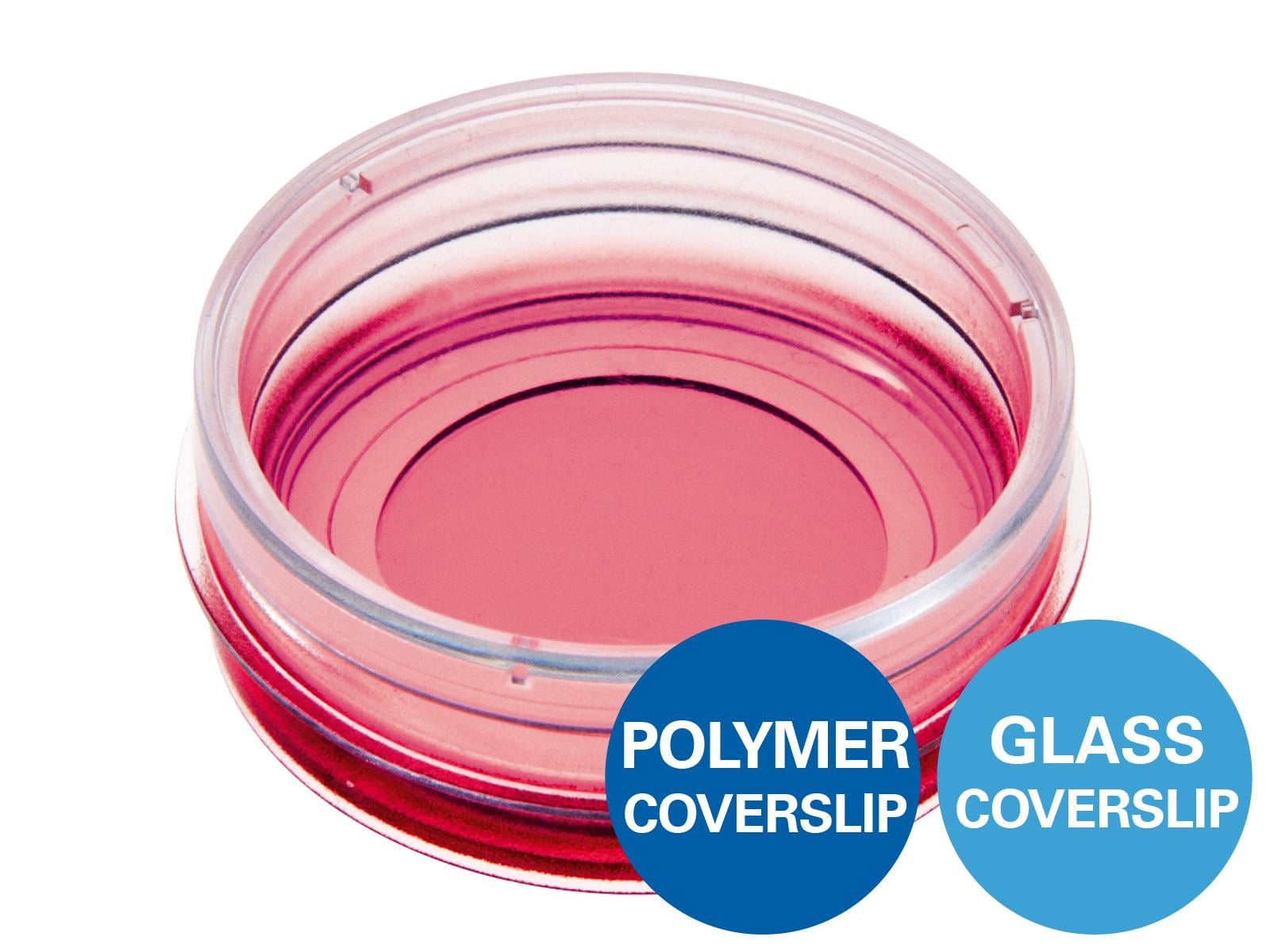
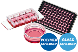
FRAP experiments require stable live cell conditions, high optical quality, and reliable fluorescence imaging. ibidi µ-Slides, and µ-Plates support live cell FRAP experiments with excellent optical quality through the ibidi Polymer Coverslip and the ibidi Glass Coverslip. For longer recovery measurements, the ibidi Stage Top Incubator helps maintain stable temperature, humidity, and CO₂ conditions.
Common Problems and Troubleshooting
Why is my fluorescence recovery weak?
A weak recovery can indicate a low mobile fraction, poor fluorescent labeling, or insufficient signal intensity.
Solution: Improve labeling quality, check expression levels, optimize exposure settings, and make sure the fluorescent signal is strong enough before bleaching.
Why is the fluorescence recovery too fast to measure?
Very fast molecular diffusion may occur before enough post-bleach images are captured.
Solution: Increase acquisition speed, reduce the time interval between images, and use a smaller bleaching region of interest.
Why is the fluorescence recovery very slow or incomplete?
Slow or incomplete recovery can result from a large immobile fraction, strong molecular binding, phototoxicity, or stressed cells.
Solution: Check cell viability, reduce bleaching intensity if possible, optimize live cell conditions, and compare recovery behavior across multiple cells.
Why does fluorescence decrease during recovery imaging?
Ongoing photobleaching during time-lapse acquisition can reduce fluorescence intensity and distort the recovery curve.
Solution: Lower laser power, reduce exposure time, increase imaging intervals where possible, and correct for acquisition bleaching during analysis.
Why is there no clear FRAP recovery curve?
An unclear recovery curve can result from poor focus, low signal-to-noise ratio, an unsuitable ROI size, or sample movement.
Solution: Use a stable imaging setup, improve focus, choose an appropriate ROI size, and ensure the sample remains stationary during acquisition.
Why do FRAP results vary strongly between cells?
High variability can be caused by biological heterogeneity, inconsistent bleaching settings, different expression levels, or variable ROI sizes.
Solution: Standardize bleaching power, ROI size, acquisition settings, and analysis parameters. Use sufficient replicates and compare similar cells or regions.
Why do cells move during FRAP acquisition?
Cell movement may occur during longer time-lapse measurements or when environmental conditions are unstable.
Solution: Maintain stable temperature, humidity, and CO2 conditions, use suitable live cell imaging chambers, and shorten acquisition time where possible.
FAQs
What is FRAP microscopy used for?
FRAP microscopy is used to study molecular mobility, diffusion, protein dynamics, and molecular exchange in living cells. It is commonly applied in membrane protein studies, cytoskeletal research, intracellular transport analysis, and live cell imaging experiments.
What is the principle of FRAP?
In FRAP, a defined fluorescent region is photobleached with high-intensity light. Fluorescence recovery is then measured over time as unbleached molecules move into the bleached area. The recovery curve provides information about molecular mobility and diffusion behavior.
What can be measured with FRAP?
FRAP can be used to estimate recovery half-time, mobile fraction, immobile fraction, and diffusion-related parameters. These values help researchers compare how molecules move under different biological or experimental conditions..
Which microscope is used for FRAP?
FRAP is commonly performed with a confocal fluorescence microscope because it allows precise laser bleaching of a defined region of interest. However, FRAP can also be performed with widefield, spinning disk confocal, or other fluorescence microscopy systems if controlled photobleaching and time-lapse imaging are possible.
References
Ishikawa-Ankerhold, H.C.; Ankerhold, R.; Drummen, G.P.C. Advanced Fluorescence Microscopy Techniques—FRAP, FLIP, FLAP, FRET and FLIM. Molecules, 2012; 17(4):4047-132. doi: 10.3390/molecules17044047
Lippincott-Schwartz, J. et al. The Development and Enhancement of FRAP as a Key Tool for Investigating Protein Dynamics, Biophys J 2018, 115(7):1146-1155. doi: 10.1016/j.bpj.2018.08.007
Axelrod, D., Koppel, D.E., Schlessinger, J., Elson, E. and Webb, W.W. Mobility measurement by analysis of fluorescence photobleaching recovery kinetics. Biophys. J., 1979;16(9):1055-69 doi: 10.1016/S0006-3495(76)85755-4

Article written by Stefanie Kiderlen, PhD
ibidi GmbH | July 2, 2026
Cell biologist and microscopy specialist with expertise in advanced imaging, live cell imaging, and 3D cell culture models. Stefanie received her PhD at the Biophysical Department at LMU, Munich, focusing on atomic force microscopy in 2D and 3D cell culture systems.