


Differential Interference Contrast (DIC)
Application
DIC is a more expensive, label-free microscopy technique with a high sensitivity to thin cellular material, even when it is located within thick tissue. It is useful for rendering contrast in transparent samples and gives brilliant pseudo-3D relief shading images. Although DIC images look very appealing, the pseudo-3D effect might be misleading in some cases, making it seem that the cells have structures that they do not have. As an example, areas inside a living cell with a different refractive index, like vacuoles and chromatin, appear as bumps, which is actually an optical impression.
Principle
Similar to phase contrast, DIC microscopy is a contrast-enhancing technique. DIC uses polarized light to convert phase delays into intensity changes (contrast). The effect is called differential, because contrast is created only in neighboring areas. Unlike in phase contrast, the DIC image is not built globally over the entire image. Instead, adjacent structures with different refractive indices are contrasted when in close contact with each other.
DIC is less sensitive to meniscus formation than phase contrast. However, DIC needs low birefringence of the microscopy chamber and the lid, making it incompatible with standard polystyrene cultureware.
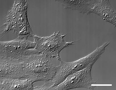
DIC microscopy of Rat1 cells. Scale bar 20 µm.
DIC Compatibility of Different Materials
| Compatible |
|
* µ-Slide I Luer 3D, µ-Slide III 3D Perfusion, µ-Slide Spheroid Perfusion
| Not compatible |
|
ibidi Solutions:
|  |