


Inverted and Upright Microscopy:
Difference, Principle, and Applications
Inverted and upright are two common microscope setups used in cell biology, live cell imaging, and fixed-sample analysis. The key difference is the position of the objective lens: inverted microscopes observe samples from below, while upright microscopes observe them from above. This affects sample preparation, sterility, incubation compatibility, and the type of labware required. Inverted microscopy is especially useful for imaging living adherent cells in culture vessels, whereas upright microscopy is often used for fixed samples, tissue sections, and larger specimens. Choosing the right setup helps improve image quality, cell viability, and experimental reliability.
Inverted Microscopy Setup
In an inverted microscopy setup, the objective lens is located beneath the specimen, pointing upward through the coverslip bottom, ideal for live cell imaging, as cells settle on the coverslip and can be observed from underneath under sterile conditions. |  |
Upright Microscopy Setup
 | In an upright microscopy setup, the objective lens is positioned above the specimen, pointing downward. This is commonly used for fixed samples, tissue sections, or in vivo imaging. |
Comparison: Inverted vs Upright Microscopy
| Feature | Inverted Microscopy | Upright Microscopy |
| Objective position | Below the sample | Above the sample |
| Best for | Live cell imaging, adherent cells | Larger samples, in vivo and tissue imaging |
| Sterility | Easier to maintain | Higher contamination risk in live cell setups |
| Typical labware | Ideal for coverslip bottom labware | Open-well labware or Petri dishes for direct access, or cell culture flasks for low-magnification microscopy |
| Incubation compatibility during imaging | Very good e.g., ibidi Stage Top Incubators | More limited |
What Is the Principle of Inverted Microscopy?
In an inverted microscope, the objective is positioned below the stage, pointing upward. For a transmitted light path, the light source and condenser are positioned above the stage, directing light downward.
The specimen is observed through the bottom of the culture vessel. For optimal imaging, the vessel bottom must match the objective’s specifications and meet the highest optical standards, as provided by the #1.5 Polymer Coverslip and the #1.5H Glass Coverslip
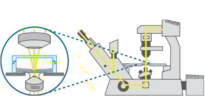
Schematic of an inverted microscope. Note the thin vessel bottom, through which the cells are observed.
When to Use an Inverted Setup?
Inverted microscopy is the preferred configuration for a wide range of live cell imaging applications as well as for imaging fixed and stained samples. Especially for live cell imaging, this setup offers distinct advantages in maintaining physiological conditions, ensuring sample integrity, and enabling precise experimental manipulation. It is especially valuable in cell biology, pharmacology, and tissue engineering, where high-resolution imaging over extended periods is essential.
Key benefits of using an inverted microscope include:
- Optimal focal plane – Adherent cells naturally settle and spread along the vessel bottom, aligning within a single focal plane for consistent imaging.
- Cell culture conditions – Inverted setups allow for larger medium volumes above the cells, improving nutrient availability and supporting long-term viability.
- Accessibility from the top – Allows medium exchange, drug application, or micropipette manipulation during imaging.
- Maintained sterility – The objective remains below the coverslip bottom, avoiding direct contact with the sample and thereby minimizing contamination risk.
- Seamless integration with incubation systems – Supports maintenance of physiological conditions during long-term live cell imaging.
ibidi Solutions for Inverted Microscopes
- μ-Slides, μ-Dishes, and μ-Plates: Incorporate a thin coverslip bottom, either a #1.5 Polymer Coverslip or a #1.5H Glass Coverslip
- ibidi Stage Top Incubators: Maintain physiological conditions during imaging
What Is the Principle of Upright Microscopy?
In an upright microscope, the objective is positioned above the stage, pointing downward toward the specimen. For a transmitted light path, the light source and condenser are positioned beneath the stage, directing light upward. The sample is typically viewed from above, either through the lid of a petri dish, directly onto a coverslip, or (when using dipping objectives) immersed in a matching medium without a coverslip.
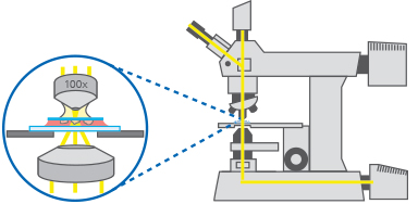
Schematic of an upright microscope. Note that the cells are squeezed between the slide and a coverslip.
When to Use Upright Microscopes
In cell biology, upright microscopes are primarily used for low-resolution phase contrast microscopy. While they can be adapted for high-resolution live cell imaging (particularly with dipping objectives) this setup is less common for cell culture applications due to the increased contamination risk and limited compatibility with incubation systems. As a result, upright configurations are more commonly used for fixed cells, tissue sections, and larger specimens, including intact tissues and in vivo imaging in small animals, often in conjunction with advanced techniques such as multiphoton microscopy.
Upright-Compatible ibidi Labware
- 3 | 8 | 12 Well Chamber Slides, removable: Silicone chamber on top of a microscopy slide can be removed after cultivation and staining for coverslip mounting
- μ-Slide VI – Flat: Designed for both upright and inverted microscopes without compromising optical integrity
FAQs
What is the difference between inverted and upright microscopy?
In inverted microscopy, the objective lens is located below the sample and images through the bottom of the vessel. In upright microscopy, the objective is above the sample and images from the top.
When should I use an inverted microscope?
An inverted microscope is best for live cell imaging, especially with adherent cells in culture. It is ideal for time-lapse experiments, sterile workflows, and studies that require controlled environmental conditions.
When should I use an upright microscope?
An upright microscope is commonly used for fixed samples, tissue sections, larger specimens, and in vivo imaging. It is well suited for slide-based workflows and applications that do not require live cell conditions.
Which microscope is better for live cell imaging?
Inverted microscopy is generally better for live cell imaging because it allows observation through the coverslip bottom while maintaining sterility and stable environmental conditions.
Do I need special labware for inverted microscopy?
Yes, inverted microscopy requires labware with an optical-quality bottom. The thickness and material of the bottom must match the objective specifications to ensure optimal image quality. All ibidi labware is designed with optimized bottom thickness and high optical quality to ensure compatibility with standard microscope objectives and reliable imaging results.

Article written by Stefanie Kiderlen, PhD
ibidi GmbH | October 13, 2025
Cell biologist and microscopy specialist with expertise in advanced imaging, live cell imaging, and 3D cell culture models. Stefanie received her PhD at the Biophysical Department at LMU, Munich, focusing on atomic force microscopy in 2D and 3D cell culture systems.