


TIRF Microscopy:
Principle, Advantages, and Applications
Total internal reflection fluorescence (TIRF) microscopy is a fluorescence imaging technique used to visualize cellular events near the glass-sample interface. By exciting fluorophores only within a thin optical section, TIRF reduces out-of-focus background and improves the signal-to-noise ratio. This makes it especially useful for live cell imaging of membrane dynamics, cell adhesion, endocytosis, exocytosis, receptor-ligand interactions, and single-molecule events. Because TIRF mainly captures structures close to the coverslip, it is best suited for adherent cells and surface-near processes rather than deep structures inside a specimen.
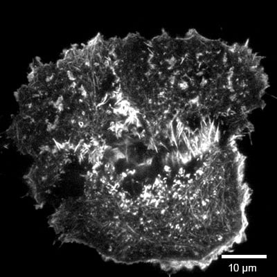
TIRF image of MCF7 cells stably transfected with LifeAct for visualizing the F-actin skeleton, captured using a 100x oil immersion objective. Image courtesy of Lena Prange, AG Wedlich-Söldner, Institute of Cell Dynamics and Imaging, University of Münster, Germany.
What Is TIRF Microscopy?
In TIRF microscopy, excitation light is directed through the glass bottom at an oblique angle. This contrasts with conventional widefield or confocal fluorescence microscopy, where the light is directed along the optical axis. Once the angle of incidence exceeds the critical angle, the light no longer propagates into the aqueous sample. Instead, it is reflected back into the glass. This reflection creates a very thin excitation zone just above the glass surface, called the evanescent field. The evanescent field rapidly decreases in intensity with increasing distance from the glass, so that only fluorophores located close to the coverslip-facing cell membrane are efficiently excited. This reduces fluorescence background from deeper regions of the cell and improves contrast for events at or near the basal plasma membrane.
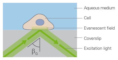
Principle of TIRF microscopy. Laser light is totally reflected at the glass-water interface, generating a thin evanescent field that selectively excites fluorophores close to the cell membrane.
When to Use TIRF Microscopy
TIRF microscopy is most useful when the biological region of interest is located close to the coverslip-facing cell membrane. It is commonly used for imaging endocytosis, exocytosis, membrane receptor behavior, vesicle docking and fusion, focal adhesions, cytoskeletal structures near the membrane, and single-molecule dynamics. TIRF typically illuminates fluorophores within roughly 60–200 nm of the interface, depending on optical conditions. It is not suitable for thick samples, suspended cells far from the surface, or experiments that require information from the full cell volume.

Want to image not only at the surface but deeper into the sample?
Learn more about confocal microscopy.
How Does TIRF Work?
TIRF microscopy is based on the refractive-index difference between the glass coverslip and the aqueous sample medium. The light travels from glass, which has a higher refractive index (n=1.52), toward the aqueous cell culture medium, which has a lower refractive index (n=1.33). At this interface, it is refracted away from the normal. If the illumination angle is increased beyond a so called “critical angle”, the light can no longer propagate into the aqueous sample. Instead, it is completely reflected back into the glass. This effect is called total internal reflection.
Although the excitation light itself is reflected, a field of light is generated on the sample side of the glass-sample interface. This field is called the evanescent field. It does not travel deeply into the sample and decreases rapidly with distance from the glass surface. Typically, it reaches only about 100–200 nm into the specimen, depending on the illumination angle, excitation wavelength, and refractive indices of the optical media. However, this thin excitation field is sufficient to excite fluorophores located close to the coverslip surface, for example near the basal plasma membrane of adherent cells. Fluorophores deeper inside the cell are not efficiently excited, which reduces background fluorescence and improves contrast for membrane-proximal processes.
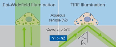
Comparison of conventional fluorescence illumination (left) and TIRF microscopy (right). In TIRF, oblique excitation light travels from the glass coverslip with a higher refractive index (n₁) toward the aqueous sample medium with a lower refractive index (n₂). At the glass–sample interface, the light is totally reflected, generating a thin evanescent field that selectively excites fluorophores close to the coverslip-facing cell membrane.
Why is the coverslip material and quality important when using TIRF microcopy?
Because the evanescent field is formed directly at the glass-sample interface, the quality of the glass bottom is critical for reliable TIRF imaging. A precise glass thickness, high optical quality, low autofluorescence, and a flat imaging surface help maintain stable illumination conditions, reduce background, and support accurate focusing with high numerical aperture objectives.
What objectives can be used for TIRF?
To generate total internal reflection, the objective must be able to direct light into the coverslip at very large angles. Therefore, high numerical aperture objectives are required, and oil immersion objectives are commonly used in objective-based TIRF systems.
What light source is ideal for TIRF and why?
Laser light is particularly beneficial for TIRF microscopy because it provides a focused, high-intensity, and collimated excitation beam.
How does the oblique illumination occur?
In TIRF microscopy, the excitation beam is directed through the objective in an off-axis position. This causes the light to enter the coverslip at an oblique angle instead of traveling straight through the optical axis. By adjusting this angle, the illumination can be moved from standard fluorescence excitation to total internal reflection.
Comparison: TIRF vs Widefield and Confocal Microscopy
TIRF, widefield fluorescence, and confocal microscopy are all fluorescence imaging methods, but they differ in excitation volume, optical sectioning, and the type of biological question they are best suited for.
| Method | TIRF | Widefield | Confocal |
| Best for | Membrane-proximal live cell events | Fast overview imaging | Optical sections and 3D structures |
| Imaging plane | ~60–200 nm near coverslip | Whole illuminated sample | Thin focal plane |
| Main advantages | Very low background and high signal-to-noise ratio | Simple, fast, sensitive | No out-of-focus light for volumetric imaging and z-sectioning |
| Main limitations | Only surface-near structures | High out-of-focus background | Slower and more phototoxic than widefield in many live cell setups |
Use TIRF microscopy when the biological process of interest occurs close to the coverslip-facing plasma membrane, such as vesicle trafficking, receptor dynamics, endocytosis, exocytosis, or cell adhesion.
Use widefield fluorescence microscopy when fast image acquisition, high sensitivity, and simple live cell imaging are more important than optical sectioning, especially in thin samples or cell monolayers.
Use confocal microscopy when you need optical sectioning, z-stacks, or 3D information from thicker samples, tissues, spheroids, or structures located deeper inside the cell up to ~100 µm.

ibidi Solutions for TIRF Microscopy
For reliable TIRF microscopy, the imaging surface is critical because the evanescent field is generated at the glass–sample interface. High-quality glass supports stable illumination, low background, and precise focusing with high numerical aperture objectives. ibidi Glass Coverslip Bottom labware is well suited for TIRF, super-resolution, and single-molecule microscopy. This #1.5H glass bottom with 170 µm ±5 µm thickness supports demanding high-resolution imaging applications.
For live cell TIRF experiments, researchers can choose from µ-Dishes, µ-Slides, and µ-Plates with a #1.5H Glass Coverslip Bottom. For long-term imaging, these formats can be combined with ibidi Stage Top Incubators to control temperature, CO₂, O₂, and humidity on the microscope.
What Are the Main Applications of TIRF Microscopy in Cell Biology?
TIRF microscopy is widely used in cell biology and biophysics to study dynamic processes close to the cell plasma membrane, such as:
Receptor–Ligand Interactions
TIRF microscopy is used to study receptor binding, receptor clustering, lateral movement, and early signaling events at the plasma membrane. Because the excitation is restricted to the near-surface region, membrane-associated fluorescence can be observed with reduced intracellular background.
Cell Adhesion and Focal Adhesions
TIRF microscopy is well suited for studying cell adhesion because focal adhesions form close to the glass-facing membrane. It can be used to analyze adhesion formation, maturation, turnover, and cell–matrix interactions in living cells.
Endocytosis, Exocytosis, and Vesicle Trafficking
TIRF microscopy enables live cell imaging of vesicle docking, fusion, internalization, and clathrin-mediated endocytosis close to the plasma membrane. This makes it useful for tracking dynamic membrane trafficking events with high contrast.
Viral Attachment and Entry
TIRF microscopy can be used to visualize fluorescent viral particles as they bind to the cell surface, move along the plasma membrane, or enter the cell. It is particularly useful for single-particle studies of early infection events.
Single-Molecule Imaging
By reducing background fluorescence, TIRF microscopy supports the detection and tracking of low-abundance fluorescent molecules near the coverslip surface. This makes it useful for single-molecule and near-membrane dynamics studies.
Advantages and Limitations of TIRF Microscopy
TIRF microscopy offers several important advantages for imaging fluorescent signals close to the cell membrane. It reduces out-of-focus fluorescence and therefore provides a high signal-to-noise ratio. It is well suited for live cell imaging of membrane-proximal processes such as endocytosis, exocytosis, receptor dynamics, vesicle trafficking, and cell adhesion.
The main limitation of TIRF microscopy is its restricted imaging depth. Since only fluorophores close to the glass–sample interface are excited, TIRF is not suitable for deep intracellular structures, thick samples, tissues, or volumetric imaging.
Common Problems and Troubleshooting
Why is my TIRF signal weak?
The fluorophores might be too far away from the glass-sample interface, or the illumination angle is not optimized for TIRF excitation.
Solution: Check cell attachment, focus position, fluorophore brightness, laser intensity, and TIRF angle. Use high-quality glass bottom labware and ensure that the structure of interest is close to the coverslip-surface.
Why is my background fluorescence high?
High background can occur when the system is not correctly adjusted to TIRF mode, when unbound fluorophores remain in the sample, or when the imaging surface is contaminated.
Solution: Optimize the TIRF angle, wash out unbound dye, reduce laser power, and use clean, low-autofluorescence glass bottom labware.
Why is my illumination uneven?
Uneven illumination can result from optical misalignment, laser coupling issues, or surface irregularities.
Solution: Check the optical alignment, clean optical components, use flat-field correction for quantitative analysis, and image on a flat, optically suitable glass surface.
Why do my cells detach during TIRF imaging?
Cells can detach if adhesion to the glass surface is insufficient, especially during medium exchange or long-term live cell imaging.
Solution: Optimize cell seeding density, incubation time, and surface coating. Handle medium changes carefully and maintain stable environmental conditions during imaging.
FAQs
What is the principle of TIRF microscopy?
TIRF microscopy is based on total internal reflection at the glass-sample interface. This generates an evanescent field that selectively excites fluorophores close to the coverslip surface.
What is TIRF microscopy used for?
TIRF microscopy is used to image fluorescent structures and dynamic processes close to the cell membrane, such as vesicles, receptors, adhesion sites, endocytosis, exocytosis, and single-molecule events.
Why is TIRF microscopy useful for live cell imaging?
TIRF microscopy reduces background fluorescence from deeper cell regions and provides high contrast for fast processes near the plasma membrane, making it well suited for live cell imaging.
What is the difference between TIRF and widefield fluorescence microscopy?
Widefield fluorescence microscopy excites a larger sample volume, which can increase background fluorescence. TIRF microscopy restricts excitation to the near-surface region, improving contrast for membrane-proximal structures.
Which samples are best suited for TIRF microscopy?
TIRF microscopy is best suited for adherent cells, membrane-associated processes, and fluorescent molecules located close to an optically suitable glass surface, such as the ibidi Glass Coverslip Bottom.
Can I Use the ibidi Polymer Coverslip for TIRF Microscopy?
TIRF is generally possible with the ibidi Polymer Coverslip, but we recommend using the ibidi Glass Coverslip Bottom.
References
Axelrod D. Total internal reflection fluorescence microscopy in cell biology. Traffic, 2001;2:764–774. doi: 10.1034/j.1600-0854.2001.21104.x
Mattheyses AL, Simon SM, Rappoport JZ. Imaging with total internal reflection fluorescence microscopy for the cell biologist. Journal of Cell Science, 2010, 123:3621–3628. doi: 10.1242/jcs.056218
Fish KN. Total Internal Reflection Fluorescence (TIRF) Microscopy. Current Protocols, 2022;2:e517. doi: 10.1002/cpz1.517

Article written by Stefanie Kiderlen, PhD
ibidi GmbH | June 10, 2026
Cell biologist and microscopy specialist with expertise in advanced imaging, live cell imaging, and 3D cell culture models. Stefanie received her PhD at the Biophysical Department at LMU, Munich, focusing on atomic force microscopy in 2D and 3D cell culture systems.