


Live Cell Imaging
Live cell imaging is a microscopy approach that observes living cells over time under controlled conditions, enabling the visualization of cellular structures, molecular interactions, and signaling dynamics with high temporal resolution. It plays a central role in cell biology and biomedical research by revealing how cells behave in real time.
To perform these experiments, researchers use carefully controlled environmental workflows that maintain temperature, CO₂, O₂, and humidity on the microscope while optimizing acquisition settings to minimize phototoxicity. These assays are essential for investigating cell motility, morphology, intracellular trafficking, cytoskeletal dynamics, and reporter activity under normal and perturbed conditions.
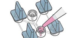
ibidi supports these applications by offering optimized labware, such as Stage Top Incubators, that simplify live cell imaging workflows and ensure excellent imaging results. Key products include Channel Slides for experiments under flow, the µ-Slide Chemotaxis for gradient-based migration, the Culture-Insert 2 Well for wound healing assays, and the µ-Slide 15 Well 3D for matrix-based angiogenesis and 3D cell culture etc.
ibidi Products
for Live Cell Imaging
Choose the optimal products for your live cell imaging experiment.
ibidi Blog Article |
Ever considered how humidity affects your live cell imaging? Delve into our blog articles "The Invisible Player: Humidity in Live Cell Imaging" and "5 Reasons Why You Should Perform Live Cell Imaging" to uncover why this often-overlooked element is crucial for successful experiments.
Selected Publications for Live Cell Imaging
Extracellular matrix labeling with a glycan-binding fluorophore was analyzed using the µ-Slide 8 Well high Glass Bottom and Collagen Type I for live cell imaging workflows.
Fiore A, Yu G, Northey JJ, et al. Live imaging of the extracellular matrix with a glycan-binding fluorophore. Nat Methods. 2025;22:1070–1080. doi:10.1038/s41592-024-02590-2.
Read article
Colorectal cancer cell migration in 3D collagen gradients was analyzed using the µ-Slide Chemotaxis for single-cell tracking.
Tetrick MG, Emon MAB, Doha U, et al. Decoupling chemical and mechanical signaling in colorectal cancer cell migration. Sci Rep. 2025;15:4952. doi:10.1038/s41598-025-89152-4.
Read article
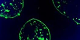
Peroxisomes were visualized in living cells using the µ-Slide 4 Well Glass Bottom for confocal live cell imaging.
Korotkova D, Borisyuk A, Guihur A, et al. Fluorescent fatty acid conjugates for live cell imaging of peroxisomes. Nat Commun. 2024;15:4314. doi:10.1038/s41467-024-48679-2.
Read article
Protein cluster formation before condensation was quantified by real-time live cell fluorescence microscopy using the µ-Dish 35 mm, high Glass Bottom.
Lan C, Kim J, Ulferts S, et al. Quantitative real-time in-cell imaging reveals heterogeneous clusters of proteins prior to condensation. Nat Commun. 2023;14:4831. doi:10.1038/s41467-023-40540-2.
Read article
Branching dynamics in pancreas-derived organoids were monitored using the ibidi Stage Top Incubator and the µ-Slide 2 Well for confocal imaging.
Randriamanantsoa S, Papargyriou A, Maurer HC, et al. Spatiotemporal dynamics of self-organized branching in pancreas-derived organoids. Nat Commun. 2022;13:5219. doi:10.1038/s41467-022-32806-y.
Read article
Chemotactic migration of human neutrophils was tracked by live cell imaging using the µ-Slide Chemotaxis and the ibidi Stage Top Incubator.
Hundhammer T, Gruber M, Wittmann S, et al. Paralytic Impact of Centrifugation on Human Neutrophils. Biomedicines. 2022;10(11):2896. doi:10.3390/biomedicines10112896.
Read article
Find more publications in the ibidi Reference Database.
Application Notes for Live Cell Imaging
Live Cell Imaging Videos


Live Cell Imaging Webinars

Live Cell Imaging White Paper
Frequently Asked Questions
What is live cell imaging and why is it used?
Live cell imaging means watching living cells over time in conditions that keep them healthy. It lets you see cell–cell interactions, migration, division, organelle transport, and signaling as they happen. Because you follow the same cells over time, you can quantify rates and trajectories with confidence. Keeping conditions stable on the microscope stage (temperature, CO₂, O₂, humidity) is critical for reproducible results.
Why is a Stage Top Incubator important?
Cells are sensitive to changes in temperature, CO₂, O₂, and humidity, which can alter behavior and image quality. A Stage Top Incubator maintains these parameters directly on the microscope, reducing evaporation, pH shifts, and focus drift during long recordings. Consistent conditions improve viability and reduce variability across positions and time points.
How can I minimize phototoxicity and photobleaching?
Keep light exposure as low as possible. Use a sensitive camera, short exposure times, and longer gaps between frames. Choose fluorophores that match your filter sets, prefer longer excitation wavelengths when you can, and illuminate only the region you need. Use antifade reagents and imaging labware with low autofluorescence to keep a good signal to noise at low light. Run small pilot tests to find the minimum light dose that still answers your question.
Which microscopy techniques are suitable for live cell imaging?
Use label free assays like wound healing, chemotaxis, and tube formation with phase contrast or DIC to see cells without staining. When you need molecule specific information, use fluorescence methods such as spinning disk confocal, multiphoton, light sheet, TIRF, or suitable super resolution techniques. Choose the method that best fits your question by balancing speed, light exposure, imaging depth, and detail. Hardware autofocus and a motorized stage help you image many fields consistently over long times.
What labware is recommended for reliable live imaging?
All ibidi products are suitable for live cell imaging and engineered for excellent optical performance with high NA objectives. µ-Slides, µ-Dishes, and Multiwell Plates with #1.5 or #1.5H coverslip bottoms provide uniform fields of view and low autofluorescence. You can choose different labware depending on your application.
How should I choose frame rate, magnification, and experiment duration?
Match the sampling to the speed of the process. Use seconds for fast events like Ca²⁺ signals, minutes for migration or proliferation, and hours for long observations. Lower magnification (4× to 10×) gives a larger field of view for population statistics; higher magnification shows subcellular detail. Make sure your computer and storage can handle the data volume. Keep light exposure low and confirm that your settings do not change cell behavior.
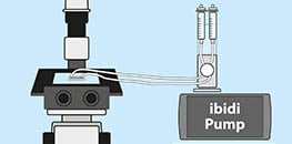
Can live cell imaging be combined with flow, gradients, or immunofluorescence?
Yes. Channel Slides with the ibidi Pump System support imaging under defined shear stress, and the µ-Slide Chemotaxis provides stable gradients for directed migration. You can fix the sample after live imaging and perform immunofluorescence, as long as the fixatives and dyes are compatible with the labware and matrix. Live reporters add dynamic readouts, but always balance time resolution with cell health.