The Invisible Player: Humidity in Live Cell Imaging
ibidi Blog | October 24, 2023 | Abhishek Derle, ibidi GmbH
Introduction
Picture this: you’ve spent countless hours meticulously setting up your live cell imaging experiment. The cells are in their prime, the microscope is ready to roll, and you’re eager to delve into the fascinating world of cellular dynamics. But hold on a minute! Before you enter the cold dark room to embark on this cellular adventure, there’s one often overlooked factor that can significantly influence the outcome of your experiment: humidity. Yes, this invisible companion to your cells plays a pivotal role in your live cell microscopy experiment. In this blog article, we’ll explore the importance of humidity and why you should give it the attention it deserves. And if you stick with us, we’ll also throw in some humidity solutions for successful live cell imaging.
Before we dive into humidity’s role in live cell imaging, let’s take a moment to appreciate the complexity of the dance of life happening within each cell. Cells are like busy little factories, where molecular machinery orchestrates fundamental processes such as cell division, signal transduction, and organelle function. Situated at the cutting edge of cell biology research, microscopic imaging techniques offer scientists a backstage pass to observe and study living cells in real-time. It gives researchers unprecedented insights into cellular processes and interactions. To conduct successful live cell imaging experiments, you must create an environment that supports cell viability and physiological functioning. This includes controlling factors such as temperature, pH, and humidity.
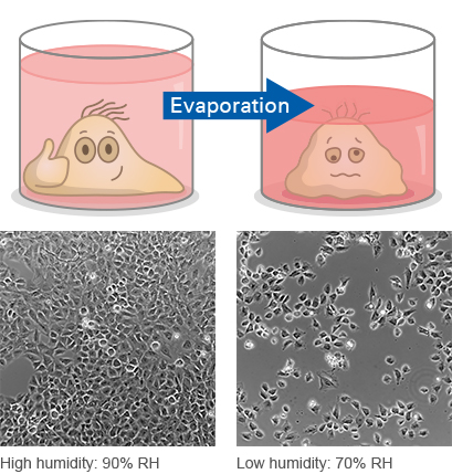
Just like the Goldilocks zone, where planets find the ideal conditions for life, cells also require the right balance of humidity to thrive. Too little, and they’ll dry out faster than a raisin in the sun. Too much, and they become waterlogged and uncooperative. The optimal humidity level typically falls within the range of 90–95% relative humidity, creating a cozy environment for your cells. Maintaining this balance is essential for live cell microscopy because it ensures that the conditions inside the culture dish closely mimic the cells’ natural habitat. This, in turn, leads to more accurate and reliable results.

Rat fibroblast cells cultured in full growth medium at different levels of relative humidity (RH) 48 hours after seeding. Phase contrast microscopy, 20x objective lens.
The Impact of Humidity on Microscopy
Now, you might be wondering how humidity affects microscopy. Well, the microscope is like the director’s chair, complete with a camera. It needs the right conditions to capture the best scenes. Here’s how humidity influences the action:
1. Cell Behavior
Changes in humidity levels can affect cell morphology, motility, and even gene expression. Cells, submerged in a cozy layer of medium, are highly sensitive to the surrounding osmotic conditions. When humidity is insufficient, water can evaporate from the cell culture medium or buffer, leading to increased solute concentration and reduced osmolality. This sets off a chain reaction of osmotic effects, which causes alterations to the ion concentrations. Such variations can disrupt numerous cellular processes, including transport across cell membranes, protein folding, and gene expression.
2. Data Reproducibility
In scientific research, reproducibility is paramount. Inconsistent humidity conditions between experiments could introduce unwanted variability into your results. It’s like trying to bake the perfect cake, but with a faulty oven. By controlling humidity, you will increase the chances of obtaining consistent and reproducible data, which makes it easier to draw meaningful conclusions from your experiments.
3. Optical Distortions
Humidity influences the optical quality of live cell images. Low humidity levels can trigger changes in the ion concentration within the cell culture medium, leading to fluctuations in the medium’s refractive index, and creating optical distortions. Your images might start to resemble an abstract painting in an art gallery—all wavy and surreal. On the other hand, high humidity can result in condensation, which produces water droplets that can obstruct the optical path and impact imaging quality.
4. Evaporation
Cells are remarkably sensitive, and even minor environmental changes can throw them off balance. When humidity levels are low, trouble brews. Any reduced humidity can trigger water evaporation from the cell culture medium, leaving behind excessive concentrations of salts and minerals. This toxic buildup then becomes a threat to the cells, often resulting in their demise.
5. Gas Exchange
Humidity doesn’t only affect the water content; it also plays a vital role in gas exchange. It has a direct impact on how gases, including oxygen, dissolve in the growth medium. When humidity levels fluctuate, it can disrupt gas solubility due to its inherent connection to pressure. In cases of insufficient humidity, gas exchange may be compromised, potentially leading to hypoxia within the cell culture. By ensuring stable humidity levels, you will provide your cells with the ability to breathe comfortably and without the need for miniature oxygen tanks!
Overall, humidity is not just a random factor to consider in live cell imaging, it’s also a critical parameter that can significantly impact the outcome of your experiments. Maintaining the right humidity levels will ensure that your cells remain healthy, behave naturally, and produce reliable and reproducible data. To achieve precise humidity control, it’s essential to employ humidity management solutions.
Humidity Solutions for Successful Live Cell Imaging
1. Use a Humidity Chamber
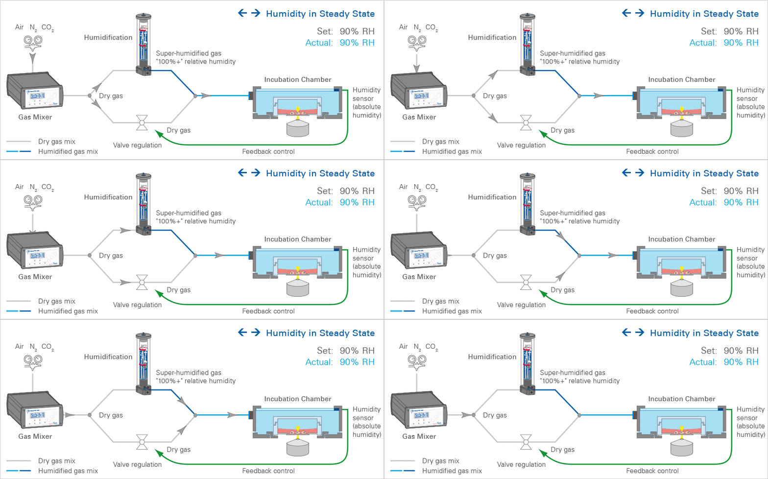
When it comes to maintaining the ideal humidity levels for your live cell imaging experiments, investing in a humidity chamber or stage top incubator is a game-changer. These purpose-built devices offer precise control over both humidity and temperature, ensuring your cells feel right at home.
Among these devices, the ibidi Stage Top Incubators really stand out. They seamlessly attach to almost every standard inverted microscope, which makes them a versatile choice. Thanks to ibidi’s patented technology for maintaining optimal humidity, your cells won’t suffer the usual negative effects that can occur during long-term assays. They are also ideal for high-resolution microscopy, as they provide maximal focus stability, and ensure that there’s no annoying condensation on the heated lid.

Learn more about all ibidi Stage Top Incubators.
2. Tight Sealing
To maintain proper humidity levels, it’s essential to achieve a tight seal on your culture dishes and chambers to prevent moisture from escaping. You can effortlessly achieve this with ibidi's ibiSeal, a self-adhesive cover film designed for sealing open wells and reservoirs in cell culture and microscopy applications. Additionally, consider using ibidi's µ-Dishes for cell culture imaging, which come equipped with a convenient lid-locking feature to further enhance humidity control.
Learn more about all ibiSeal and µ-Dishes for cell culture imaging.
3. Parafilm
Placing small strips of Parafilm onto the reservoirs/wells of the cell culture vessel will suppress the effects of evaporation.
4. Regular Monitoring
Continuously monitor humidity levels during your experiments. Invest in a hygrometer to keep a close eye on the environment. It’s worth noting that this functionality is a built-in feature of the ibidi Stage Top Systems.
Learn more about all Parameters for Live Cell Imaging.
Conclusion: Keep Your Cells Hydrated in Optimal Cell Imaging Conditions!
So, let’s raise a glass of water to humidity. This unsung hero of live cell imaging is a critical factor that profoundly influences the quality and reliability of experimental results. Maintaining optimal humidity levels within the imaging environment is essential for ensuring cell viability, preventing dehydration-induced artifacts, and facilitating accurate observations of dynamic cellular processes. Precise control of humidity not only sustains the physiological conditions necessary for cell survival, but also aids in minimizing experimental variability and enhancing the reproducibility of findings, mitigating humidity effects on cells. Researchers need to recognize the significance of humidity control as an indispensable component of live cell microscopy protocols. It enables the study of cellular behavior in a more physiologically relevant context and advances our understanding of complex biological phenomena.
 (1)
(1)  (0)
(0)


