


How to Use Calcium Imaging for Investigating Cell Signaling
ibidi Blog | May 15, 2023 | Kristen Engevik, Baylor College of Medicine, Houston, Texas, USA
Calcium is an important messenger that influences multiple cellular processes, including a cell’s ability to migrate, communicate, and respond to irritants or foreign bodies. Due to its importance in cell function, calcium inside of cells (known as intracellular calcium), is highly regulated. Calcium is primarily stored within organelles, such as the endoplasmic reticulum and mitochondria, and is released in response to specific cues. This tight regulation makes intracellular calcium an excellent tool for understanding cell function and signaling dynamics.
Calcium: A Versatile Indicator for Cellular Processes
There are several fluorescent indicators for intracellular calcium which can be used to study cell function. These indicators allow researchers to examine cellular responses during wound healing, infectious disease, drug treatment, and more.
In our lab, we use calcium signaling as a readout to understand the ability of viruses to exploit host signaling responses during infection and to dissect the signaling pathways involved. To do this, we utilize live cell imaging of cells that contain fluorescent calcium indicators where increases in fluorescence indicate increases in intracellular calcium.
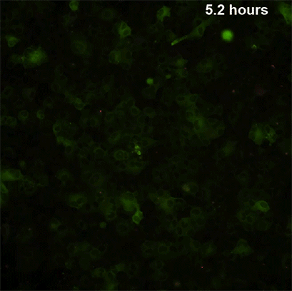
Live cell imaging of rotavirus infected African green monkey kidney cells (MA104). MA104 cells, shown in green, expressing the calcium indicator GCaMP6s, where increases in green indicate increases in intracellular calcium. In this live cell imaging video, rotavirus infected cells are in red – the Hyser lab has previously reported that during rotavirus infection, rotavirus causes an increase in intracellular calcium in infected cells and a subsequent activation of specific signaling pathways in neighboring cells that elicits increases in uninfected cells’ intracellular calcium signaling (Chang-Graham et al., 2020).
Types of Calcium Indicators and Applications
Calcium indicators exist in two main categories: chemical indicators (i.e., dyes) and genetically encoded calcium indicators (i.e., the calcium indicator is encoded into a cell line, animal model, or system). Calcium indicators have been utilized for in vivo and in vitro work using microscopy, high-throughput screening, and flow cytometry.
However, for the purpose of this article, we will focus on using microscopy to visualize and quantify calcium signaling in vitro. The type of calcium indicator used for live cell imaging experiments depends on factors like the intended cell line, length of imaging, and desired outcome for qualitative or quantitative data.
1. Intended Cell Line
For primary cell lines, the straightforward approach is to use fluorescent dyes (chemical indicators), where the fluorescent calcium indicators are simply added to cultured cells and can be imaged following an incubation period. These dyes are also an excellent approach for first-time experiments to gather preliminary data. One important tip when using fluorescent dyes is to thoroughly wash the cells before and after adding, as dyes can cause high background fluorescence, making it more difficult to focus cells during microscopy and live imaging properly.
Fluorescent dyes are typically brighter and more stable compared to genetically encoded calcium indicators. However, dyes are generally less specific than genetic sensors and can have higher background fluorescence. In live cell imaging, fluorescent dyes (including Fura-2AM, Fluo-4AM, Indo 1AM, etc.) can be effective for short term live cell imaging. A potential caveat is that in certain cells, there can be an issue with the cells retaining the dyes or not properly loading into certain cell types or subpopulations. Additionally, there is a tendency for cells to pump out the dyes over time. While the addition of anion transporter inhibitors can prevent the cells from pumping out the dye, these inhibitors can be cytotoxic to certain cell types and may complicate conditions if other treatments are being used to assess changes to calcium signaling. One important consideration with calcium dyes is whether inhibitors or agonists will be used. Inhibitors and agonists may also affect the calcium dyes’ ability to reflect expression similar to that in native tissue.
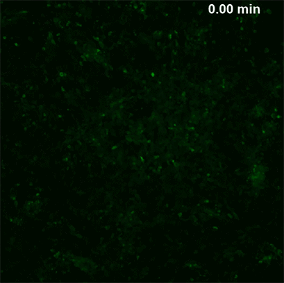
Live, short-term imaging of human intestinal organoid monolayers during agonist treatment with ADP. These organoid monolayers express the calcium indicator GCaMP6s in green. ADP is a known agonist for eliciting intracellular calcium signaling in cells. Agonist treatment is an excellent approach to testing for dose responses of treatments.
For extensive calcium signaling experiments, established cell lines (including organoids) can be transduced to express a genetically encoded calcium indicator such as GCaMPs, yellow cameleon, etc. Genetically encoded calcium indicators are integrated into the host genome, allowing cells to function normally while also including a fluorescent protein-based indicator that will transiently be expressed and reflect changes within intercellular calcium signals. Improvements to the different available genetically encoded calcium indicators have resulted in brighter indicators with superior photostability and expanded dynamic range, thus improving the calcium sensitivity. Lentiviral transduction is an excellent tool to use in cell lines that will allow stable expression of the indicator of your choice within your cell line. A potential issue of the genetically encoded reporter system is that not all cell lines are easily transduced.
2. Length of Imaging
Timing is everything. With live cell imaging, the length and intervals of time are important parameters to keep in mind. The main concern during imaging is photobleaching and phototoxicity. Continuous application of the excitation light should be avoided, and it’s important to select an exposure time and light power that is strong enough to detect calcium signaling but not overpowering where it causes damage to cells. The length of imaging will rely on the experiment. For testing agonists or cellular responses, this is typically a shorter imaging run of 5-20 minutes, capturing images every 1-2 seconds. For experiments studying calcium signaling during wound repair or viral infection, we image over the course of 18-20 hours, capturing images every minute.
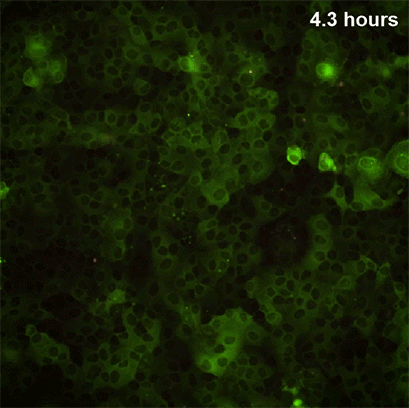
Live cell imaging of rotavirus infected African green monkey kidney cells (MA104). MA104 cells, shown in green, expressing the calcium indicator GCaMP6s, where increases in green indicate increases in intracellular calcium. In this live cell imaging video, rotavirus infected cells are in red – the Hyser lab has previously reported that during rotavirus infection, rotavirus causes an increase in intracellular calcium in infected cells and a subsequent activation of specific signaling pathways in neighboring cells that elicits increases in uninfected cells’ intracellular calcium signaling (Chang-Graham et al., 2020)
General Tips for Live Cell Imaging
It’s important to consider the cell line to be used. For studying calcium dynamics (or other cellular signals), using a cell line that forms as a monolayer will ensure the regions of interest will remain in focus to capture signaling dynamics for the best quality of video and clean quantifying. For experiments involving cell to cell communication and calcium dynamics, cell line confluency is also an important consideration. For best quality imaging, it is important to select the appropriate slide. There are cell cultures that prefer glass versus optical grade plastic slides, so it is best to test the chamber slides. We use ibiTreat µ-Slide 8 Well high chambered coverslip for our cell lines.
For live cell imaging, it is important to plate cells at a density that allows a confluent monolayer to form but does not cause the cells to overcrowd and clump. One final tip is using the proper media during imaging. Many traditional cell medias contain phenol red which can cause a high background fluorescence. This can make it difficult to separate true signal from background noise during imaging. Therefore, it is important to wash cells with PBS and replace the media with a phenol red free culture media or commercially available media intended for live cell imaging.
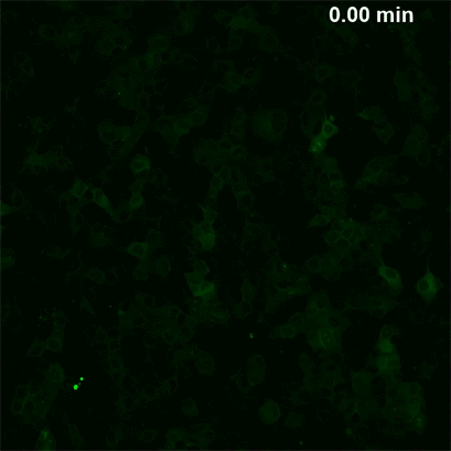
Live, short-term imaging of African green monkey kidney cells (MA104) before and after Thapsigargin treatment. MA104 cells, shown in green, expressing the calcium indicator GCaMP6s, where increases in green indicate increases in intracellular calcium. Thapsigargin is a well-known inhibitor for the sarco/endoplasmic reticulum Ca2+-ATPase (which is important for sequestering calcium from the cytosol). Through this inhibition, Thapsigargin induces endoplasmic reticulum stress and can result in increased intracellular calcium originating from the endoplasmic reticulum.
References:
Chang-Graham AL, Perry JL, Engevik MA, Engevik KA, Scribano FJ, Gebert JT, Danhof HA, Nelson JC, Kellen JS, Strtak AC, Sastri NP, Estes MK, Britton RA, Versalovic J, Hyser JM. Rotavirus induces intercellular calcium waves through ADP signaling. Science. 2020 Nov 20;370(6519):eabc3621. doi: 10.1126/science.abc3621.
Read article
Dr. Kristen Engevik, PhD
Dr. Kristen Engevik, PhD, is a postdoctoral fellow in the lab of Dr. Joseph Hyser at Baylor College of Medicine, Houston, Texas. Her research focuses on elucidating host molecular signaling associated with rotavirus infection to understand the mechanistic consequences of rotavirus dysregulation of host pathways. She has an extensive background using live cell imaging microscopy to study gastrointestinal epithelial cells and their responses to damage and infection.
As an avid microscopist, you can see her work posted on her Twitter (@thekengevik) or on Instagram (@the_engevik_labs).

 (2)
(2)  (0)
(0)