


Spheroids | Organoids | 3D Cell Culture
Enhance Your 3D Cell Culture
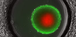
3D cell cultures, such as spheroids, organoids, or cells in 3D scaffolds, or microengineered tissue models, such as organ-on-a-chip models, are increasingly adopted for in vitro studies, delivering a more precise representation of the in vivo environment compared to traditional 2D cultures. And now, with advanced cultivation methods such as dynamic flow culture or bioprinting, it is possible to create 3D tissue conditions that are even closer to the biological origin. Hence, 3D cell culture models and advanced cultivation methods lead to more physiologically relevant results with regards to cell viability, gene expression, disease modeling, and drug testing, as well as toxicity studies. They also support the 3R principles by providing animal-free alternatives (Replace), reducing the number of required animal studies (Reduce), and enabling more physiologically relevant in vitro assays (Refine).
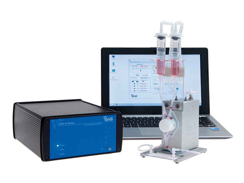
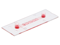
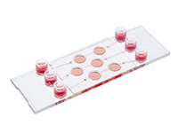
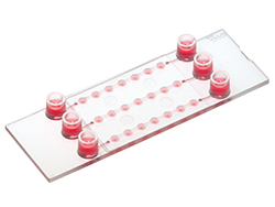
ibidi provides advanced, customizable solutions for the creation and image-based analysis of 3D cell models. Our modular systems are designed to simplify the complexity underlying cutting-edge 3D cell-based assays. By integrating various components of the ibidi products, researchers can effortlessly manage complex designs, enhancing the robustness and efficiency of their scientific investigations. With the ibidi µ-Slides / µ-Dishes / µ-Plates, the ibidi Pump System, or the ibidi Stage Top Incubators, you can take your research to the next level and achieve more accurate and reliable results.
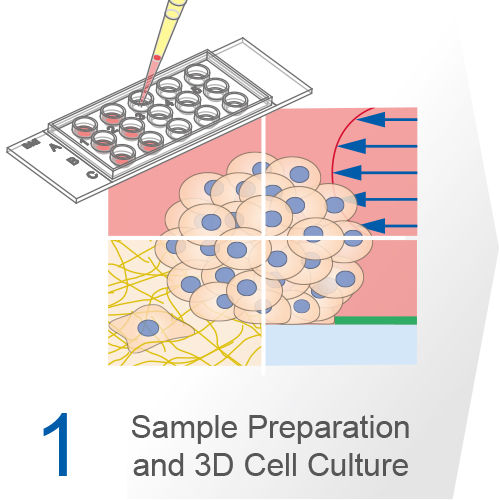
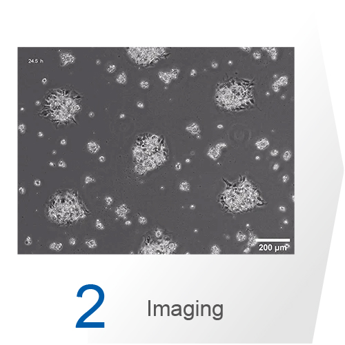
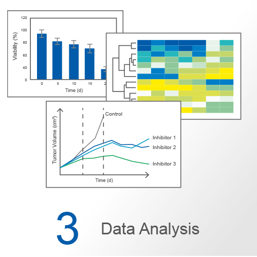
Experimental Workflow of a 3D Cell Culture Assay
Discover the solutions for all your experimentation needs, from sample preparation to 3D cell cultivation, imaging, and analysis in the 3D Cell Culture Workflow chapter.
3D Cell Culture Systems and Applications
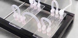
3D cell culture experiments require robust setups that work with absolute precision and reliability. The ibidi products, including the µ-Slides / µ-Dishes / µ-Plates, the ibidi Pump System, and the ibidi Stage Top Incubators, are designed for performing and analyzing 3D cell culture assays under physiologic conditions. These modular components can be combined to build complex 3D models, including automated long-term 3D cell culture and microfluidic organ-on-a-chip systems under precisely controlled flow conditions.
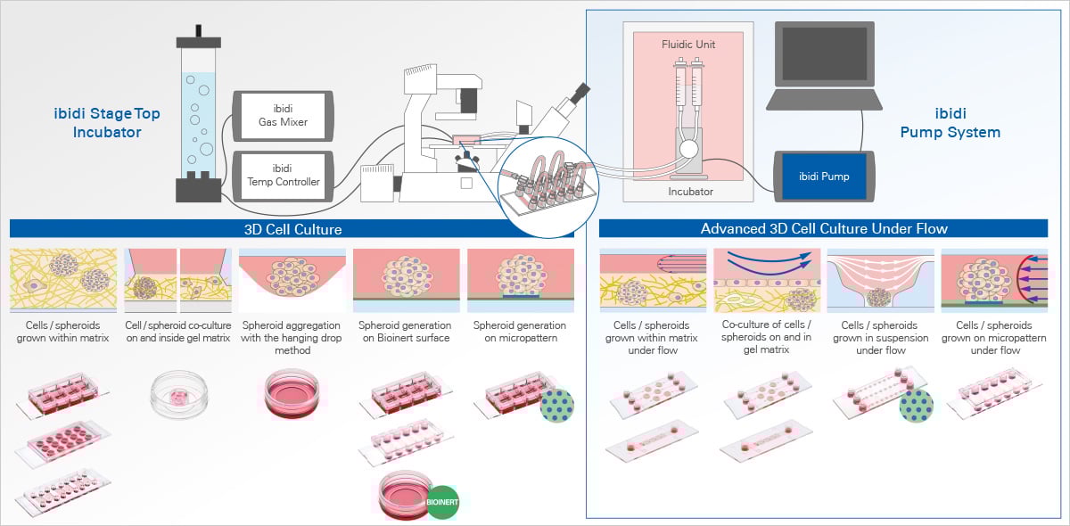
ibidi Solutions for Assays with 3D Cell Models
ibidi Blog Articles |
Explore how organoids are revolutionizing research in our latest blog article Organoids: The Miniature Labs, and get a comprehensive overview of the different systems in 3D Cell Culture Models.
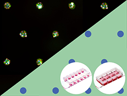
Selected Publications Using 3D Cell Culture Models
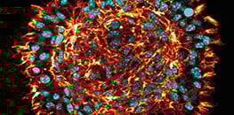
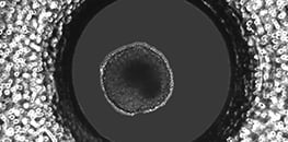
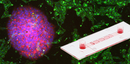
Human blastoids were cultured on a 3D matrix using the µ-Slide 8 Well.
Karvas RM, Zemke JE, Ali SS, Upton E, Sane E, Fischer LA, Dong C, Park KM, Wang F, Park K, Hao S, Chew B, Meyer B, Zhou C, Dietmann S, Theunissen TW. 3D-cultured blastoids model human embryogenesis from pre-implantation to early gastrulation stages. Cell Stem Cell., 2023, 10.1016/j.stem.2023.08.005.
Read article
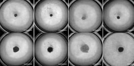
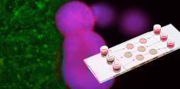
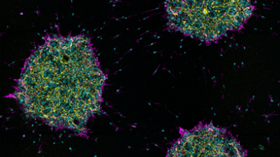
3D sandwich cultures of squameous cell carcinoma cells were done in the µ-Plate 96 Well 3D.
E. Hoque Apu, S.U. Akram, J. Rissanen, H. Wan and T. Salo. Desmoglein 3 – Influence on oral carcinoma cell migration and invasion. Experimental Cell Research, 2018, 10.1016/j.yexcr.2018.06.037.
Read article
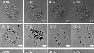
The 12 Well Chamber, removable was used for the cultivation of 3D human acinar ductal metaplasia cells.
da Silva, L., Jiang, J., Perkins, C. et al. Pharmacological inhibition and reversal of pancreatic acinar ductal metaplasia. Cell Death Discov., 2022, 10.1038/s41420-022-01165-4.
Read article
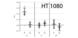
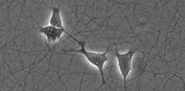
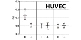
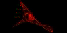
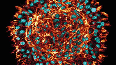
3D cell culture of human breast epithelial MCF10A cells was done in the µ-Slide 8 Well.
H. Grobe, A. Wüstenhagen, C. Baarlink, R. Grosse and K. Grikscheit. A Rac1-FMNL2 signaling module affects cell-cell contact formation independent of Cdc42 and membrane protrusions. PloS one, 2018, 10.1371/journal.pone.0194716.
Read article
Scientific 3D Cell Culture Posters
Presented at the International Cell Culture Under Flow Meeting 2024 , Chicago, USA.
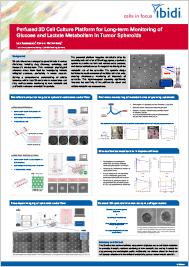
Presented at the SelectBio Conference Organoids and Spheroids Europe 2024, Rotterdam, The Netherlands.
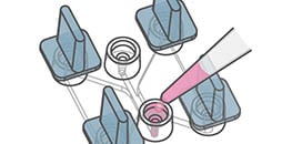
3D Cell Culture User Protocol

Philipp's Trick:
What’s an easy way to seal and perfuse 3D bioprinted structures?
Find out on our Tips and Tricks page!
3D Cell Culture Application Notes

3D Cell Culture User Protocols
3D Cell Culture Webinars


Frequently Asked Questions
What is 3D cell culture?
3D cell culture refers to growing cells in three-dimensional structures such as spheroids, organoids, hydrogel matrices, scaffolds, or microengineered tissue models. In contrast to conventional 2D monolayer culture, cells can interact with neighboring cells and extracellular matrix components in all spatial directions. This often results in more physiologically relevant morphology, differentiation, and cellular behavior. 3D cultures are therefore useful for studying complex biological processes under conditions that more closely resemble tissue-like environments.
Which 3D cell culture models can be used?
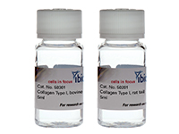
Common 3D cell culture models include spheroids, organoids, cells embedded in extracellular matrix gels, scaffold-based cultures, and perfused organ-on-a-chip systems. The ideal model depends on the biological question, cell type, assay complexity, imaging requirements, and desired throughput. For matrix-based assays, biomaterials such as Collagen Type I are commonly used to create defined 3D environments.
What is the difference between spheroids, organoids, and organ-on-a-chip systems?
Spheroids are simple, compact 3D cell aggregates, often generated from established cell lines or primary cells, and commonly used for tumor biology, cell viability, invasion, and drug response studies. Organoids are more complex, self-organizing 3D structures, typically generated from stem cells or primary tissue-derived cells, and are used to model organ development, tissue function, or disease. Organ-on-a-chip systems combine living cells with microfluidic channels to recreate physiological conditions such as perfusion, mechanical stimulation, or tissue barriers, making them useful for advanced disease modeling and tissue interaction studies.
How can 3D cell cultures be imaged?
3D cell cultures can be analyzed using phase contrast, fluorescence microscopy, confocal microscopy, and other high-resolution imaging methods. Reliable imaging depends on optical quality, low autofluorescence, and compatible vessel geometry. ibidi µ-Slides, µ-Dishes, and µ-Plates are designed for microscopy-based workflows, allowing cultivation, staining, and imaging in the same vessel to reduce handling and sample disturbance.
Can live cell imaging be performed with 3D cell cultures?
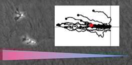
Yes. 3D cell cultures can be monitored by live cell imaging to study spheroid growth, organoid development, invasion, migration, morphogenesis, or treatment responses over time. For long-term experiments, stable temperature, CO2, humidity, and oxygen control are important. ibidi Stage Top Incubators help maintain these conditions directly on the microscope.
Can 3D cell culture be performed under flow?
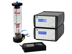
Yes. Combining 3D culture with perfusion improves nutrient delivery, waste removal, and physiological relevance. ibidi offers solutions such as the µ-Slide I Luer 3D, µ-Slide III 3D Perfusion, and µ-Slide Spheroid Perfusion, which can be combined with the ibidi Pump System to create defined flow conditions for matrix-based cultures, spheroids, and long-term perfusion experiments.
What are the applications of 3D cell culture?
3D cell culture is widely used in cancer biology, stem cell research, tissue engineering, regenerative medicine, toxicology, immunology, and drug discovery. Typical applications include tumor spheroid assays, invasion studies, organoid research, vascular biology, and advanced disease models. These models help bridge the gap between simplified 2D assays and complex in vivo systems by providing more biologically relevant in vitro conditions.
Is 3D cell culture suitable for high-throughput screening?
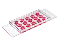
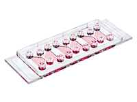
Yes. 3D cell culture can be adapted for high-throughput workflows using standardized multiwell formats such as the µ-Plate 96 Well 3D. This makes it easier to perform compound screening, toxicity testing, and image-based phenotypic analysis while maintaining improved biological relevance compared to conventional 2D assays.