


The Surfaces and Coatings of the ibidi Chambers
Find Out More
Contact us for a printed version or download the posters "Microscopy With ibidi" or "Choosing the Best Microscopy Labware" as PDF.
ibidi Surfaces and Coatings
Growth, development, and signaling of cultured cells strongly depend on the used surface. ibidi's μ-Slides, μ-Dishes, and μ-Plates have a thin coverslip bottom (ibidi Polymer Coverslip or Glass Coverslip, both with excellent optical quality). They are available with several surfaces. The ibidi μ-Slides, μ-Dishes, and μ-Plates that have an ibidi Polymer Coverslip bottom can be coated in a similar process to standard plastic labware, while fully retaining image quality. ibidi offers Collagen I, Collagen IV, and Poly-L-Lysine coatings.

ibidi Polymer Coverslip

Glass Coverslip
 ibiTreat (Tissue Culture-Treated) Surface Excellent adhesion of adherent cells | 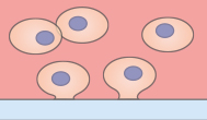 Hydrophobic, Untreated Surface Weak adhesion of adherent cells, application of specific coatings | 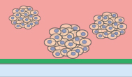 Bioinert Surface No adhesion of adherent cells |
 µ-Patterned Surface Spatially defined adhesion of adherent cells on spots, different spot geometries available | 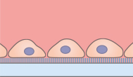 Coated Surface Culture of adherent cells on a Collagen IV or Poly-L-Lysine surface |
 Glass Surface Adhesion of adherent cells (coating might be required), special microscopy applications |
ibiTreat
Excellent cell adhesion
- Culture of adherent cells
- ECM coatings possible
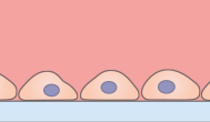
The tissue culture-treated (TC-treated), hydrophilic ibiTreat surface provides excellent cell adhesion, even without a defined protein coating. However, ECM protein coatings can be done on ibiTreat without any restrictions. The ibiTreat surface is ideal for culture of adherent cells that do not need any specific coating.
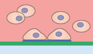
Untreated
Weak cell adhesion
- Culture of adherent cells
- ECM coatings possible
The hydrophobic Untreated surface provides weak cell adhesion, if not previously coated with an ECM protein. ECM protein coatings can be done on the Untreated surface without any restrictions. The Untreated surface is ideal for the culture of adherent cells that require a specific coating.
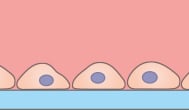
Bioinert
No cell adhesion
- Culture of suspension cells, cell aggregates, spheroids, embryoid bodies (EBs), organoids
- ECM coatings not possible
The hydrophilic Bioinert surface hinders any protein attachment, thus inhibiting subsequent cell attachment. Unlike with the ibiTreat and Untreated surfaces, a coating is not possible. The Bioinert surface is ideal for the culture of suspension cells and cell aggregates.
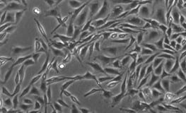
Application Example: Cultivation of Adherent Cells on Different Surfaces
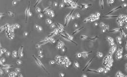
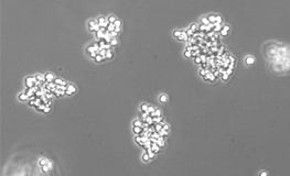
On the ibiTreat surface, all adherent cell types show a strong cell adhesion. A weaker cell adhesion is observed on the Untreated surface since there is only a serum protein coating. On the Bioinert surface, cells do not attach at all and spheroid formation can be observed in most of the cell types. Depending on the cell-cell-adhesion of the specific cell type, spheroids are formed more or less homogeneously.
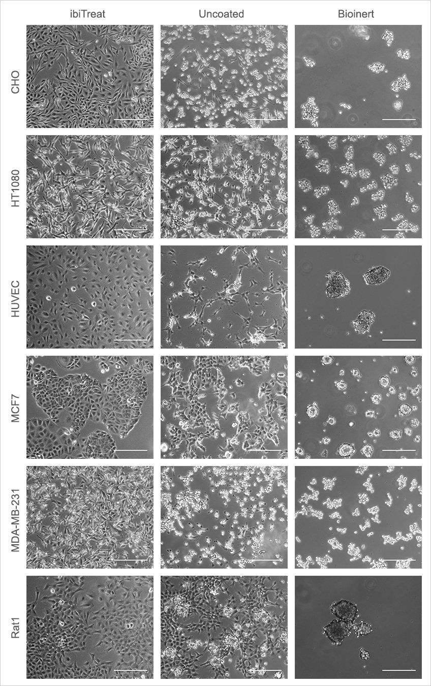
Different adherent cell types were seeded on the ibiTreat, Untreated, and Bioinert surfaces, respectively. The cells were cultured in full growth medium without prior ECM protein coating. Images were taken 24 hours after seeding. Phase contrast microscopy, Scale bar is 300 µm.
ibidi Surfaces Show no Autofluorescence
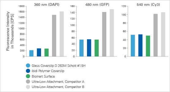
Both the flat, thin bottom material and the excellent optical quality of the ibidi Polymer Coverslip enable high-resolution microscopy without any disruptive autofluorescence.