Wound Healing and Cell Migration
Impedance Measurements
The ECIS Wound Healing Assay replaces the traditional “scratch” assay. Instead of disrupting the cell layer mechanically with a pipette tip and then following the migration of cells with a microscope, the ECIS system employs electric signals to both wound and monitor the healing process.
Experimental Example: Healing Function of Serum
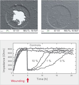
Electrical wounding is directed to a small population of cells in contact with the 250 μm electrode thus creating a well defined wound.
After wounding, the healthy cells around the electrode immediately migrate and replace the dead cells on the electrode. Depending on the fetal calf serum (FCS) concentration, the original plateau is once again reached after 10, 13, or 20 hours.
For further information:
Keese CR, Wegener J, Walker SR, Giaever I. Electrical wound-healing assay for cells in vitro. PNAS, 2004, 101(6):1554-9.
Read article
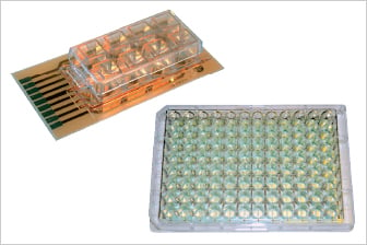
Cell migration measurements via automated wound healing are performed using the following electrode arrays:
|
8 well format: |
96 well format: |
Optical Measurements
Optical measurements of wound healing and cell migration can easily be taken with ibidi’s Culture-Insert, which provides two culture reservoirs each separated by a 500 μm wall. Culturing cells in both reservoirs and then removing the insert, results in two well-defined cell patches. An assay-specific Image Analysis can then be used to generate quantitative data based on image data acquired using video microscopy.