


The oxygen (O2) concentration at normal atmospheric pressure is around 20–21%. However, the O2 levels in healthy body tissues, and in our blood, are much lower. Variations in the oxygen concentration are part of the normal body system, with ranges from 14% in the alveoli to 5% in peripheral tissues.
The oxygen levels can be even lower in pathological situations (hypoxia). This especially occurs in tumor tissue, which is characterized by markedly low O2 levels that range from approximately 4% to a minimum of below 0.5%.
In live cell imaging assays, the O2 concentration is another important parameter that needs to be controlled, particularly when working with tumor cells or when analyzing the effects of hypoxia.
McKeown SR (2014) Defining normoxia, physoxia and hypoxia in tumours-implications for treatment response. Br J Radiol 87(1035):20130676. 10.1259/bjr.20130676.
read abstract
ibidi Solution
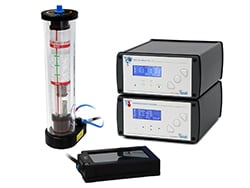
For hypoxia assays, the CO2/O2 versions of the ibidi Stage Top Incubation Systems allow for the precise control of the oxygen levels in your cell culture vessel. The ambient O2 level can be controlled within a range of 1%–21%, and can be easily adapted to the experimental requirements.
To make accurate O2 measurements, we recommend using one of the systems from ColibriPhotonics and PreSens GmbH.
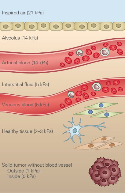
O2 Partial Pressure in Tissue
(1 kPa ≙ 1% in gas mix ≙ 7.5 mm Hg)
The in vivo O2 concentration in most types of tissue is around 2–5 kPa, which corresponds to 2–5% oxygen in air.
Read on and learn more about how to prevent Condensation in live cell imaging.