


Immunofluorescence Using ibidi Labware
ibidi provides several labware solutions for immunofluorescence staining: Chambered Coverslips, Channel Slides, Chamber Slides, removable, µ-Dishes, and µ-Plates. The right format depends on the microscopy setup, reagent volume, handling requirements, mounting method, and whether short-term or long-term sample storage is needed.
Comparison of ibidi Labware for Immunofluorescence
| Feature | Chambered Coverslips | Channel Slides | Chamber Slides, removable |
|---|---|---|---|
| Product example | µ-Slides | Channel Slides | Chamber Slides, removable |
| Bottom material | Glass Coverslip or Polymer Coverslip | Glass Coverslip or Polymer Coverslip | Standard glass slide |
| Additional coverslips required? | No | No | Yes, for mounting |
| Microscope type | Inverted | Inverted | Inverted and upright |
| Mounting medium | Non-hardening | Non-hardening | Hardening |
| Sample storage | Short-term | Short-term | Long-term |
| Typical use | Parallel immunofluorescence assays without coverslip handling | Low-volume immunofluorescence staining and exact medium exchange | Mounted samples for long-term storage or staining jar workflows |
Benefits of ibidi µ-Chambers
- High-resolution imaging
The ibidi slides are ideal for widefield fluorescence, confocal imaging, FRAP, FRET, FLIM, and undisturbed phase contrast imaging. - Fast and simple handling
The all-in-one chambers simplify your immunofluorescence protocol. - Cost-effective experiments
Only a small number of cells and low reagent volumes are needed.

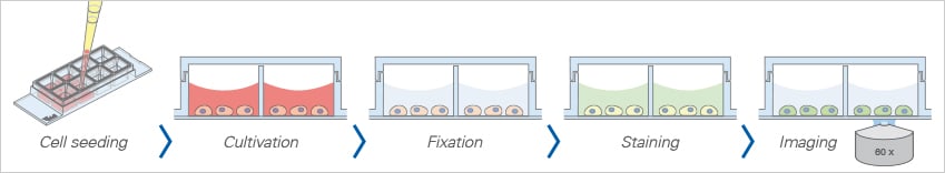
Comparison of Immunocytochemistry Protocols: Traditional Staining vs. Staining With ibidi Solutions
When using any of the ibidi solutions, the immunofluorescence staining protocol is much shorter than the traditional protocol. There is no need to grow the cells on loose coverslips—the cells can be grown and stained directly in the ibidi slides.
Protocol With Cells on Coverslips
Traditional method with nail polish mounting
- Sterilize coverslips and slides
- Coat coverslips
- Place sterile coverslips into 6-well plate
- Seed cells in large volume
- Peel off the coverslip
- Wash
- Fix – wash – permeabilize – wash – block
- Incubate in primary antibody – wash – incubate in secondary antibody
- Wash
- Mount cells with mounting medium
- Mount coverslip with nail polish
Protocol With ibidi µ-Slides
Time-saving method using all-in-one chambers
- Sterilize coverslips and slides
- Coat coverslips
- Place sterile coverslips into 6-well plate
- Seed cells in large volume
- Peel off the coverslip
- Wash
- Fix – wash – permeabilize – wash – block
- Incubate in primary antibody – wash – incubate in secondary antibody
- Wash
- Mount cells with mounting medium
- Mount coverslip with nail polish
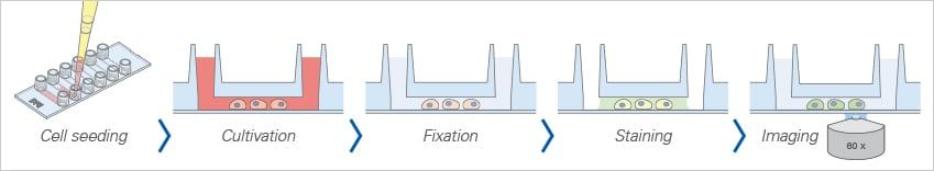
Chambered Coverslips for Immunofluorescence
When using chambered coverslips, such as the ibidi µ-Slides, the cell culture and the whole immunofluorescence staining protocol can be performed in the same well, without the necessity of any additional coverslips or tweezers. After mounting, the results can be observed through the coverslip bottom using high-resolution microscopy.
Advantages
- No coverslip handling
- Parallel assays without cross-contamination
Limitations
- Storage time is restricted to several weeks, because of the gas exchange through the plastic material from which the slides are made
Find more information and technical details about the coverslip bottom of the ibidi chambers here.
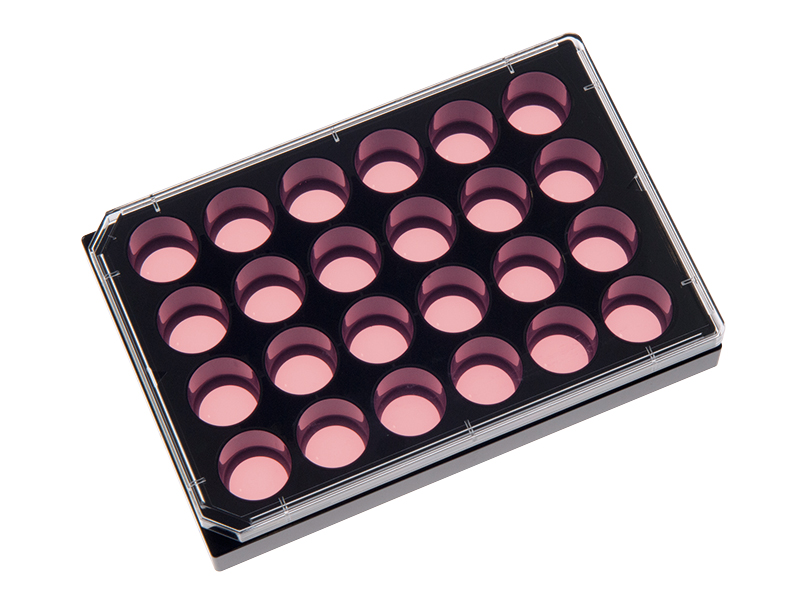
The ibidi µ-Slides are available in a variety of well numbers, volumes, and geometries that fit your specific experiment.
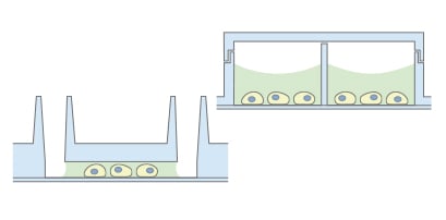
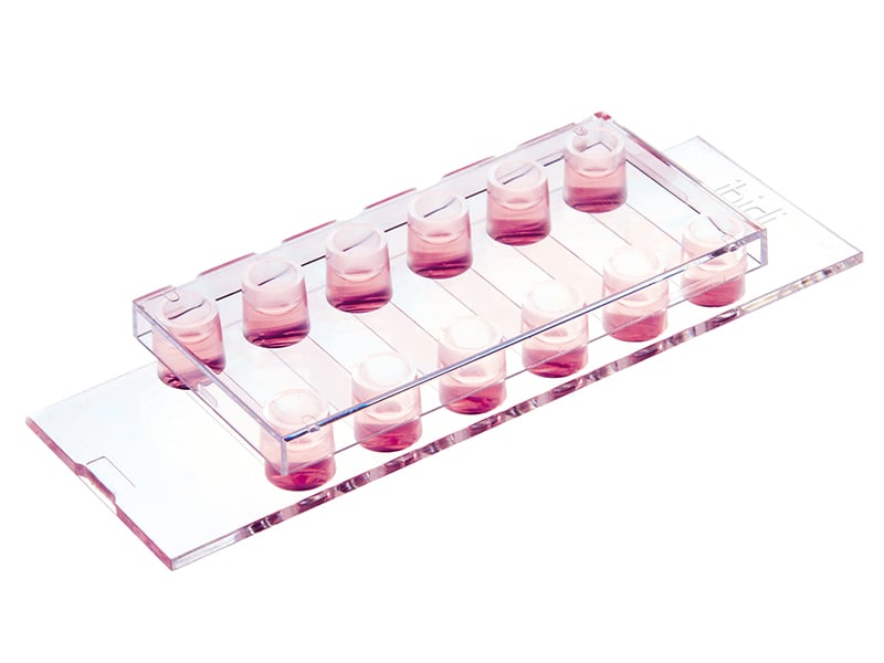
Channel Slides for Immunofluorescence
The ibidi Channel Slides are particularly suitable for immunofluorescence stainings: The geometry of the ibidi Channel Slides is ideal for the exact exchange of small medium amounts, which is necessary during immunocytochemistry stainings. In addition, the coverslip bottom of the channel µ-Slides eliminates the need for additional coverslips.
Advantages
- Meniscus-free phase contrast microscopy
- Low volumes of reagents needed
- Homogeneous cell and antibody distribution
Limitations
- Storage time is restricted to several weeks, because of the gas exchange through the plastic material from which the slides are made
Find more information and technical details about the coverslip bottom of the ibidi chambers here.
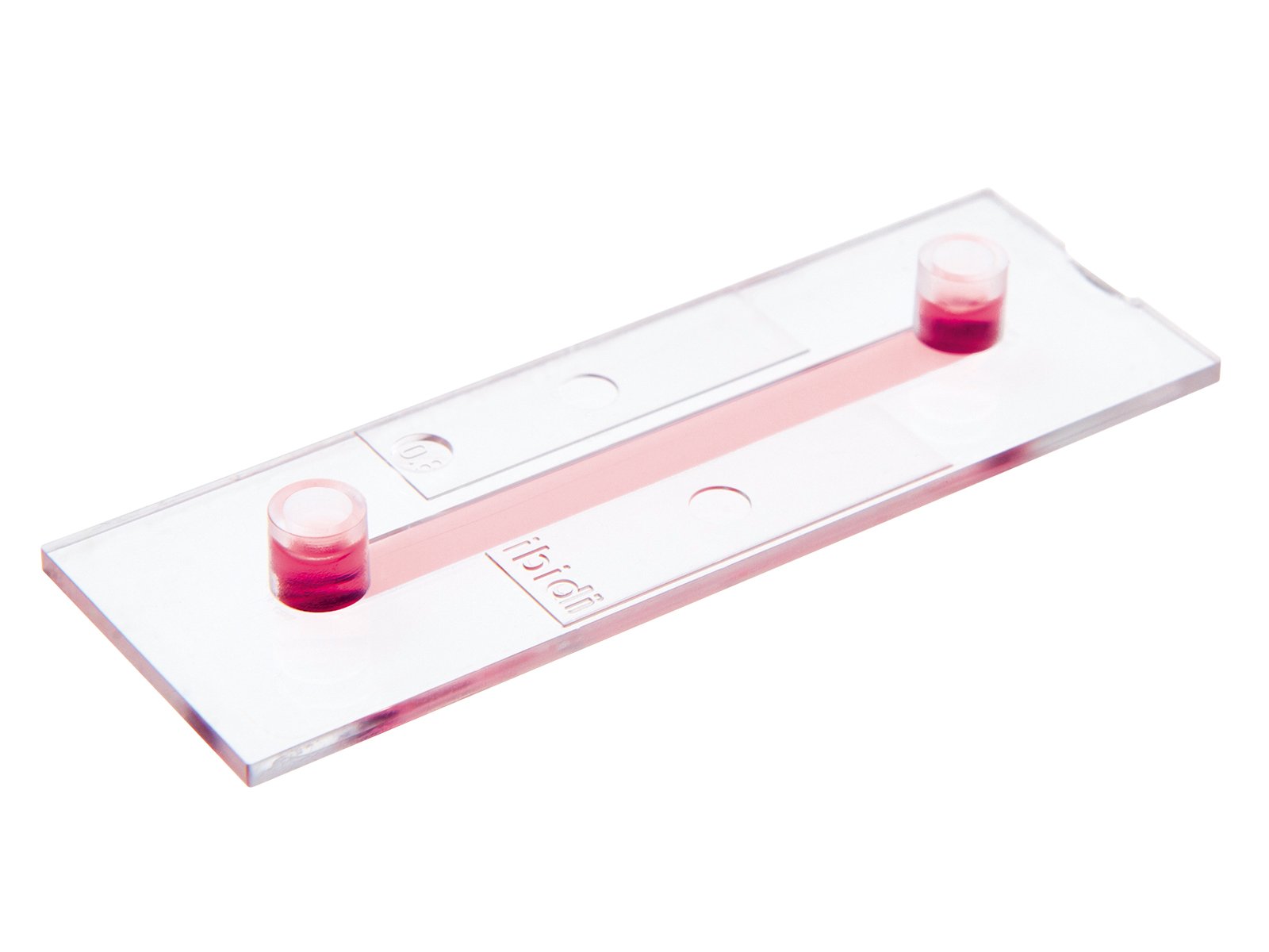
The µ-Slide VI 0.4 is ideal for general low-volume immunofluorescence assays.
The µ-Slide I Luer enables immunofluorescence staining in low-density cultures in the context of flow experiments.
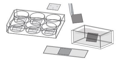
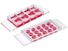
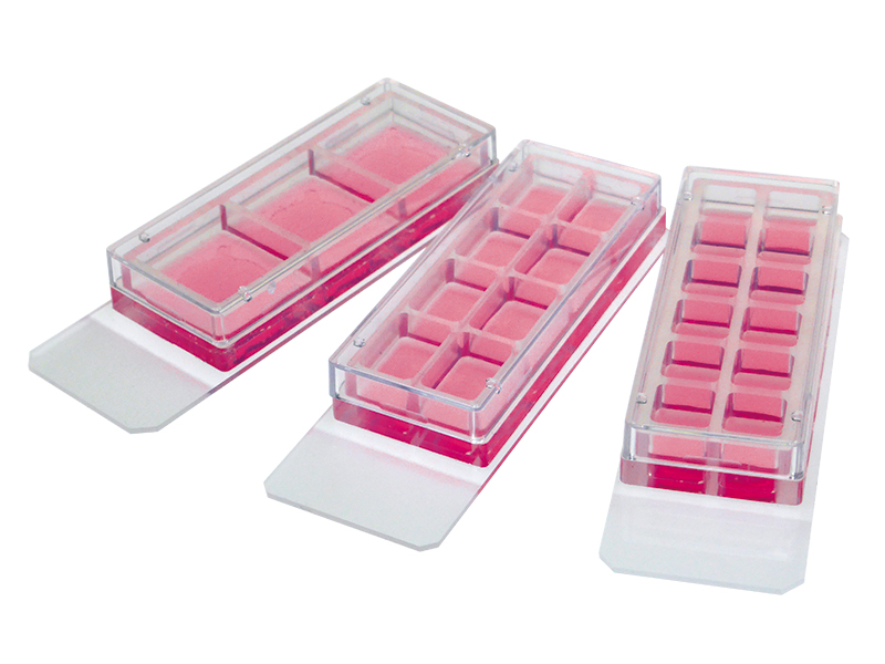
Chamber Slides, removable for Immunofluorescence
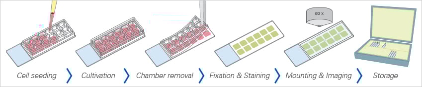
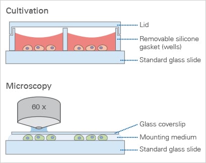
In the ibidi Chamber Slides, a silicone gasket with separated chambers is mounted on a standard glass slide. The ibidi Chamber Slides for immunofluorescence are ideal for the long-term storage of samples that are mounted with a glass coverslip.
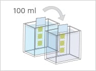
For high-throughput approaches, the silicone gasket can be removed before starting the staining. This allows for the processing of several slides at once in a staining jar. As an alternative to low-throughput experiments, the silicone gasket can be left in position, allowing for separate staining in each well.
Advantages
- Ideal for long-term storage
- High-throughput screening possible
Limitations
- No high-resolution microscopy during cell cultivation
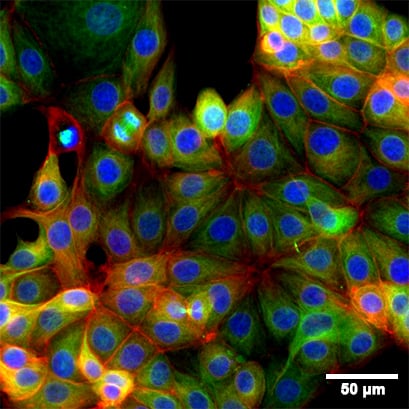
Fluorescence microscopy of immunostained MCF-7 cells in the 8 Well Chamber, removable. Green: alpha-Tubulin. Red: F-actin (phalloidin). Blue: nuclei (DAPI). Widefield fluorescence images were taken with a 20x objective.
Mounting Medium and Sample Storage
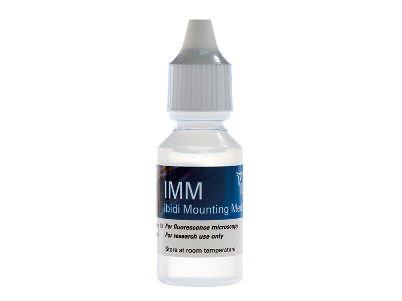
The ibidi Mounting Medium and the ibidi Mounting Medium with DAPI have a very low autofluorescence, prevent photobleaching, and allow the sample to be stored for several weeks on the µ-Slide without the need for additional coverslips.
If your experiment requires the long-term storage of immunostained samples, we recommend using the ibidi Chamber Slides, removable.
Application Notes and Protocol Resources
Find detailed immunofluorescence staining protocols in the ibidi Application Notes:
- AN 02: Fluorescence Staining using a µ-Slide I (PDF)
- AN 09: Fluorescence Staining using a µ-Slide VI 0.4 (PDF)
- AN 15: Fluorescence Staining using a µ-Slide y-shaped (PDF)
- AN 16: Fluorescence Staining Using the µ-Slide 8 Well high (PDF)
- AN 49: Fluorescence Staining using a 12 Well Chamber, removable (PDF)
- AN 50: Fluorescence Staining using a 3 Well Chamber, removable (PDF)
- AN 45: Mounting Medium Types (PDF)