


Successful Immunofluorescence Image Acquisition and Quantification
Presented by Shuntaro Yamada
Center for Translational Oral Research, University of Bergen
Course Objectives
Level: Beginner
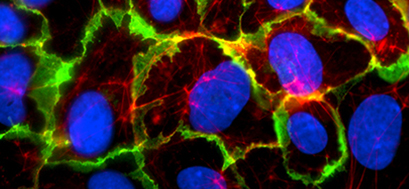
Immunofluorescence is a powerful method used to visualize structures and molecules of interest within cells and tissues. However, researchers are often challenged when trying to obtain and process sharp and brilliant images. For successful immunofluorescence images, each step—including cell preparation, staining, and image acquisition—needs to be optimized.
This ibidi online webinar is an extension of the previous webinar, How to Set Up a Successful Immunofluorescence Experiment, and is once again hosted by our invited speaker Shuntaro Yamada. During his first webinar, Yamada shared his practical tips and tricks for immunofluorescence staining as he guided us through each step of a standard immunofluorescence protocol. This follow-up webinar expands on that topic with an in-depth description of how to perform successful image acquisition and quantification.
After completing the webinar, you will have advanced knowledge and a better understanding of the necessary tools required to process beautiful immunofluorescence stainings.
Target Group
This webinar is designed for scientists and technical associates who have experience with cell culture and sterile working techniques, but want guidance when performing and troubleshooting immunofluorescence experiments.
During this webinar, we will present image and processing examples using the ImageJ software, FIJI. Therefore, it is beneficial to preinstall the software and get familiar with its basics functions.
Schedule
Topics
- Introduction to immunofluorescence staining (User Protocol 10)
- Basic image acquisition and quantification
- Discussion about your experimental aims and setups
Speaker

Shuntaro Yamada
Center for Translational Oral Research, University of Bergen
Shuntaro is a D.D.S/Ph.D. candidate at the Center for Translational Oral Research in the Tissue Engineering Group at the University of Bergen.
His research interests include the mechanical regulation of stem cell fate for bone tissue regeneration. In addition, he has extensive experience and expertise in immunofluorescent staining and imaging of cells and embryonic or postnatal tissues.
Check out Shuntaro's Instagram Channel for his beautiful immunofluorescence cell images: @stemcell_art
In preparation for this webinar, please feel free to listen—or re-listen—to Yamada’s first webinar about the basic steps of a standard immunofluorescence protocol: How to Set Up a Successful Immunofluorescence Experiment.
What Our Participants Say
"I found the ibidi Online Course Successful Immunofluorescence Image Acquisition and Quantification to be extremely helpful and informative. The instructors were very knowledgeable and approachable, and provided useful tips for successful staining. I also appreciated the practical training on image processing and analysis with Fiji (ImageJ), which really enhanced the learning experience. Overall, I would highly recommend this course to anyone looking to improve their skills in immunofluorescence staining."
Raghad Alghazali, Linköping University, Linköping, Sweden
https://liu.se/