


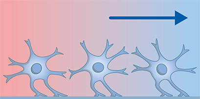
Neural Migration and Chemotaxis
During embryonic development, neural progenitor cells undergo a series of complex migration and chemotaxis events to form the different structures of the nervous system, including the brain and spinal cord. Neural progenitor cells originate in the neural tube and undergo radial migration to reach their final destination, where they differentiate into various types of neurons and glial cells. This process is tightly regulated by a variety of molecular cues, including signaling molecules and adhesion molecules, which guide the migrating cells along chemical gradients. For example, the axons of developing neurons are guided by gradients of chemoattractant and chemorepulsive signals that help them navigate toward their appropriate targets (axon guidance).
Migration and chemotaxis also play a role in the immune response of the nervous system and brain repair. After injury or disease, neurons and microglia (which are the primary innate immune cells of the brain), migrate to the site of injury to help repair damaged tissue and restore function (wound healing).
Find Out MoreFind further detailed information about the planning, conduction, and data analysis of chemotaxis assays and wound healing and migration assays in our Application Chapters. |
|

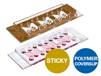
ibidi Solutions for Chemotaxis Assays
|
|
|
|
|
|
|
|
Selected References
Microglia Migration
Investigating the motility of microglial cells using the Culture-Insert 2 Well in µ-Dish 35mm.
Muth C, Hartmann A, Sepulveda-Falla D, Glatzel M, Krasemann S. Phagocytosis of Apoptotic Cells Is Specifically Upregulated in ApoE4 Expressing Microglia in vitro. Front Cell Neurosci. 2019 May 3;13:181. doi: 10.3389/fncel.2019.00181.
Read Article
Monitoring Axon Outgrowth
Live cell microscopy in the µ-Slide Chemotaxis was used to follow axon outgrowth in a 3D gel, in response to a chemical stimulus.
Terheyden-Keighley D, Brand-Saberi B, Theiss C. Real-Time Imaging of Accessible Axon Guidance Assays in Three-Dimensional Culture. Journal of Neurology and Experimental Neuroscience. 2017 January 5; 2(2): 34-39. doi:10.17756/jnen.2016-015.
Read Article
Read on and learn more about Neuronal Function and Activity, Vascularization / Angiogenesis, and Shear Stress and Barrier Function.