


Neuronal Activity
Neuronal function and activity rely on various electrical and chemical signals, which can be measured using methods like electrophysiology or calcium imaging.
Electrophysiology involves the direct measuring of electrical activity in neurons using electrodes inserted into neurons, for example, to measure its membrane potential or synaptic activity (Patch-clamp technique).
Calcium imaging is a powerful technique that allows researchers to study neuronal activity by monitoring changes in intracellular calcium concentrations. It is a valuable tool to investigate the dynamics and functional consequences of calcium signaling in neurons using sensors that change their fluorescence upon calcium-ion binding. These indicators can be loaded into neurons, and changes in calcium fluorescence can be monitored using fluorescence microscopy.
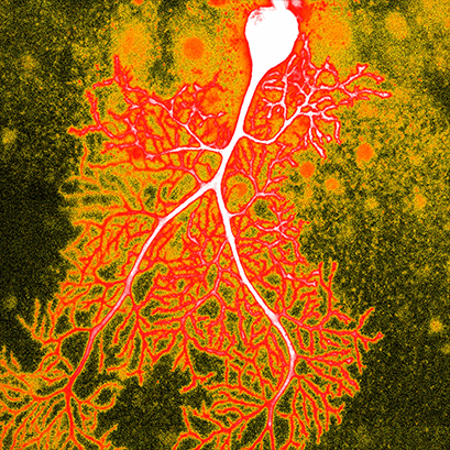
Purkinje cell from the cerebellum, visualized using confocal microscopy and calcium imaging.
Image courtesy: Dana Simmons, The University of Chicago, USA
ibidi Blog Article |
Learn How to Use Calcium Imaging for Investigating Cell Signaling in our blog article
ibidi Solutions for Neuronal Function and Activity
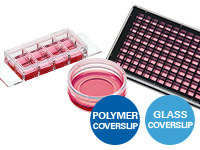
The ibidi µ-Slides and µ-Dishes include different geometries that combine optimal conditions for everyday cell culture and functional cell-based assays. They are ideal for immunofluorescence, live cell imaging, and high-resolution microscopy. The ibidi labware is available with the ibidi Polymer Coverslip and the ibidi Glass Coverslip. |
|
The ibidi µ-Plates are ideal for high throughput drug screening and large-scale knock-down and overexpression experiments with high-resolution microscopy as readout. These imaging plates are compatible with robotics and plate readers due to an ANSI/SLAS (SBS) standard format. The ibidi µ-Plates have 24, 96, or 384 wells and are available with the ibidi Polymer Coverslip and the ibidi Glass Coverslip with extremely low autofluorescence for undisturbed fluorescence microscopy. |
|
The ibidi Stage Top Incubators provide physiological conditions for live cell imaging on every standard inverted microscope. They include CO2 and O2 control (e.g., for hypoxia experiments) and actively controlled humidity. They are available for single slides and dishes along with multiwell plates. |
|

Selected References
Calcium Imaging of Neuronal Cells
The µ-Dish 35 mm, high was used for Calcium imaging to evaluate cell functionality after differentiating human induced pluripotent stem cells (iPSCs) into neurons.
Bianchi F, Malboubi M, Li Y, George JH, Jerusalem A, Szele F, Thompson MS, Ye H. Rapid and efficient differentiation of functional motor neurons from human iPSC for neural injury modelling. Stem Cell Res. 2018 Oct;32:126-134. doi: 10.1016/j.scr.2018.09.006.
Read article
Read on and learn more about Vascularization / Angiogenesis, Shear Stress and Barrier Function, and Neural Migration and Chemotaxis.