


Vascularization / Angiogenesis
The nervous system requires a constant supply of oxygen and nutrients to function properly, and the vasculature provides the infrastructure. In the central nervous system (CNS), the brain and spinal cord rely on a complex network of blood vessels to supply oxygen and glucose to the neurons and remove waste products. The blood-brain barrier (BBB), a specialized structure formed by the endothelial cells of the brain's capillaries, helps to regulate the exchange of substances between the blood and the brain to maintain a stable environment. Additionally, the brain has its own unique system of blood vessels called the cerebrovascular system, which helps to regulate blood flow and protect the brain from injury.
In the peripheral nervous system (PNS), blood vessels supply oxygen and nutrients to the peripheral nerves, which are responsible for transmitting signals to and from the CNS. The PNS also has a network of small blood vessels, which provide oxygen and nutrients to the nerve cells and protect them from injury.
After a stroke, traumatic brain or nerve injury, the essential blood flow can be disrupted, resulting in a sudden drop of oxygen (hypoxia) in the surrounding tissue. This then triggers the formation of new blood vessels from the preexisting vascular network (angiogenesis) to restore oxygen and nutrient supply to the brain. In addition, the new blood vessels deliver immune cells and growth factors to the site of injury, which promotes tissue repair and nerve regeneration.
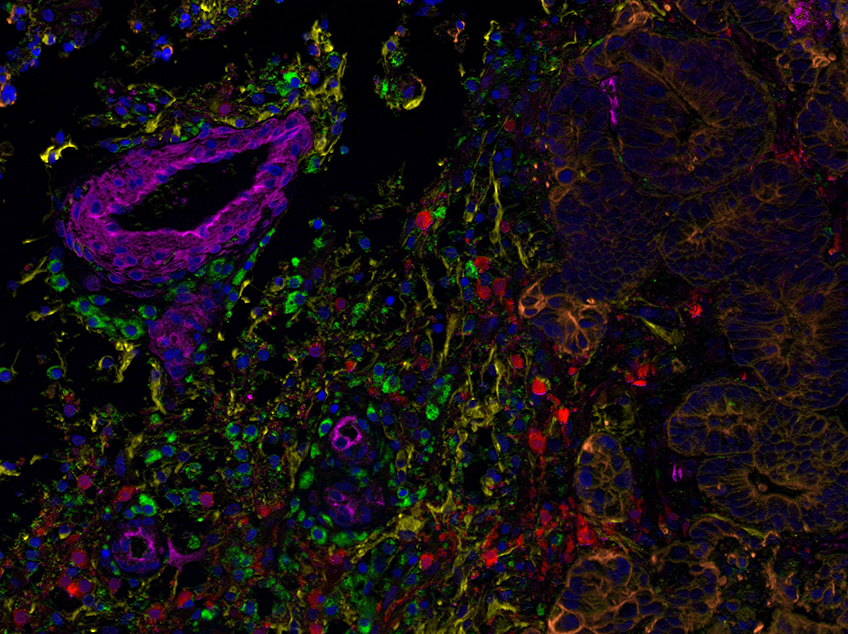
Multiplex immunofluorescence staining of a metastatic brain adenocarcinoma and its tumor environment (TME). On the right side of the image, there are tumor cells expressing cytokeratin 18 in orange (Alexa Fluor® 568). The TME is comprised of an abundant population of macrophages, visualized by CD68 expression (Alexa Fluor® 488, green) and a network of fibroblasts labeled with desmin (Alexa Fluor® 555, yellow). Also present in the TME are blood vessels expressing CD31 (Alexa Fluor® 647, pink). The granular staining shows the enzyme Aspartoacylase (Alexa Fluor® 594, red), which is expressed in neural tissue. Nuclei are counterstained with DAPI in blue.
The image was acquired using a Multispectral Microscope (Nikon 90i with a CRI multispectral camera) at 200X magnification.
Image courtesy: Sara Cascais and Mireia Castillo-Martin, MD, PhD, Champalimaud Foundation, Lisbon, Portugal
![]()
Hypoxia occurs in the central nervous system as a result of a stroke, brain or nerve injury, or during tumor growth. It can severely affect neuronal function and the effectiveness of potential treatments and drugs. Therefore, researchers should consider working under hypoxia when simulating neuronal conditions like stroke or brain cancer, as it accurately reflects the physiological oxygen levels in these conditions. The ibidi Stage Top Incubators can mimic hypoxic conditions on every inverted standard microscope during live cell imaging experiments.
Angiogenesis is also involved in the development of brain tumors. Tumors require blood supply for growth, and angiogenesis can promote the growth of new blood vessels to supply the tumor with nutrients and oxygen. Inhibiting angiogenesis can be a potential treatment strategy for brain tumors.
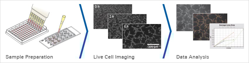
Angiogenesis can be studied in vitro with the help of tube formation assays. The assay allows the investigation of angiogenesis promoting factors or screening for angiogenesis inhibiting molecules to develop new brain cancer therapies.
Find Out MorePlease find more detailed information about the planning, conduction, and data analysis of angiogenesis assays here. |
|
ibidi Solutions for Angiogenesis Assays
|
|
|
|
|
|
|
|

Selected Reference
Tube Formation Assay to Study Vessel Regeneration After Traumatic Brain Injury
The ibidi µ-Slide 15 Well 3D was used for tube formation assays to study the role of FGF20, a member of the fibroblast growth factor family in promoting angiogenesis after traumatic brain injury.
Guo R, Wang X, Fang Y, Chen X, Chen K, Huang W, Chen J, Hu J, Liang F, Du J, Dordoe C, Tian X, Lin L. rhFGF20 promotes angiogenesis and vascular repair following traumatic brain injury by regulating Wnt/β-catenin pathway. Biomed Pharmacother. 2021 Nov;143:112200. doi: 10.1016/j.biopha.2021.112200.
Read article
Read on and learn more about Neuronal Function and Activity, Shear Stress and Barrier Function, and Neural Migration and Chemotaxis.