


Tumor Cell Migration
Cell migration occurs during the whole cascade of cancer development and is especially important during invasion, which is the initial step of metastasis. Following chemotactic migration, a cancer cell invades into the surrounding tissue and the local vasculature. This step is guided by protrusion (extension) of the cell membrane and its connection to the extracellular matrix. Migration is a precisely regulated process that can be guided by both cell-internal signals and cues of the local microenvironment. Many factors such as mechanical and chemical signals are sensed by specific cellular receptors (e.g., integrins, chemokine receptors, and growth factor receptors), thereby influencing cancer cell migration.
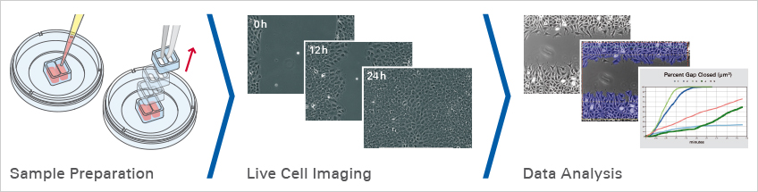
To develop therapies that prevent tumor cell migration, it is necessary to understand the mechanisms of the stimuli in tumor cells and their surrounding microenvironmental cells that lead to migration. Cell migration assays, such as the classical scratch assay or wound healing assay, give information about the speed and characteristics of cell migration (e.g., after a gene knock-down or a drug treatment). If the directed cell migration towards a chemoattractant needs to be measured, a chemotaxis assay should be conducted.
Polacheck, W. J., Zervantonakis, I. K., & Kamm, R. D. (2012). Tumor cell migration in complex microenvironments. Cellular and Molecular Life Sciences. 10.1007/S00018-012-1115-1
Read article
Find Out MoreContinue reading about the planning, conduction, and data analysis of wound healing and migration assays here. |
|


Live cell imaging of a wound healing and migration assay using the MCF7 breast cancer cell line in the Culture-Insert 4 Well. Physiological conditions were maintained with the ibidi Stage Top Incubation System. Objective lens 10x, phase contrast microscopy.
ibidi Solutions for Tumor Cell Migration Assays
|
|
The ibidi Stage Top Incubators provide physiological conditions for live cell imaging on every standard inverted microscope and are ideal for cell migration assays over longer periods of time. They include CO2 and O2 control as well as actively controlled humidity. They can be used with single dishes (e.g., the Culture-Insert 2 Well in µ-Dish 35 mm, high) as well as with multiwell plates (e.g., the Culture-Insert 2 Well 24). |
|

Selected References
Analysis of the migration of pancreatic tumor cells using the using the ibidi Culture-Inserts
Cheng, Y., Imanirad, P., Jutooru, I., Hedrick, E., Jin, U. H., Hoffman, A. R., de Araujo, J. L., Morpurgo, B., Golovko, A., & Safe, S. (2018). Role of metastasis-associated lung adenocarcinoma transcript-1 (MALAT-1) in pancreatic cancer. PLOS ONE. 10.1371/JOURNAL.PONE.0192264
Read article
Analysis of wound healing in a breast cancer model using the ibidi Culture-Inserts
Martinez-Ordoñez, A., Seoane, S., Cabezas, P., Eiro, N., Sendon-Lago, J., Macia, M., Garcia-Caballero, T., Gonzalez, L. O., Sanchez, L., Vizoso, F., & Perez-Fernandez, R. (2018). Breast cancer metastasis to liver and lung is facilitated by Pit-1-CXCL12-CXCR4 axis. Oncogene. 10.1038/s41388-017-0036-8
Read article
Migration assay with prostate cancer cells using the ibidi Culture-Inserts
Tseng, J. C., Huang, S. H., Lin, C. Y., Wang, B. J., Huang, S. F., Shen, Y. Y., & Chuu, C. P. (2020). ROR2 suppresses metastasis of prostate cancer via regulation of miR-199a-5p–PIAS3–AKT2 signaling axis. Cell Death & Disease. 10.1038/s41419-020-2587-9
Read article