


Chemotaxis
Chemotaxis of cancer cells and their associated inflammatory and stromal cells is essential during tumor progression and dissemination. It especially plays a role during the metastatic cascade, where a tumor cell invades, intravasates, extravasates, and finally grows at a distant site. Chemotaxis is tightly regulated in healthy cells and mediated by chemokines, growth factors, and their receptors. However, during cancer progression, reprogramming of the chemotactic pathways can abrogate this regulation in favor of metastasis.
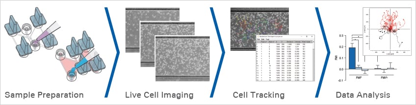
A chemotaxis assay is conducted to analyze whether or not a cell type directly orients and migrates towards a defined chemoattractant. Chemotaxis is a critical process during cancer development and immune infiltration, making chemotaxis assays a powerful tool in cancer research.
Roussos, E. T., Condeelis, J. S., & Patsialou, A. (2011). Chemotaxis in cancer. Nature Reviews Cancer. 10.1038/NRC3078
Read article
Find Out More
Continue reading about the planning, conduction, and data analysis of chemotaxis assays here.

3D live cell imaging of migrating HT-1080 cancer cells in a Collagen matrix. LifeAct-expressing HT-1080 cells (green) were seeded in a 1.5 mg/ml Collagen Type I, Rat Tail layer (white) in the µ-Slide Chemotaxis. Cell migration was documented by taking a photo every 300 seconds on a Zeiss Confocal Microscope LSM 880 AxioObserver using a water immersion objective lens 40x/1.2.
ibidi Solutions for Chemotaxis Assays
|
|
|
|
|
|

Selected References
Evaluation of the chemotactic function of NET-DNA (neutrophil extracellular traps) in breast cancer cells using the µ-Slide Chemotaxis
Yang, L., Liu, Q., Zhang, X., Liu, X., Zhou, B., Chen, J., Huang, D., Li, J., Li, H., Chen, F., Liu, J., Xing, Y., Chen, X., Su, S., & Song, E. (2020). DNA of neutrophil extracellular traps promotes cancer metastasis via CCDC25. Nature. 10.1038/s41586-020-2394-6
Read article
Analysis of the motility of single tumor cells using the µ-Slide Chemotaxis in different cancer types
Agarwal, E., Goldman, A. R., Tang, H. Y., Kossenkov, A. v., Ghosh, J. C., Languino, L. R., Vaira, V., Speicher, D. W., & Altieri, D. C. (2021). A cancer ubiquitome landscape identifies metabolic reprogramming as target of Parkin tumor suppression. Science Advances. 10.1126/sciadv.abg7287
Read article
Directed migration in cancer-associated fibroblasts (CAFs) and mesenchymal stem cells using the µ-Slide Chemotaxis
Mestre-Farrera, A., Bruch-Oms, M., Peña, R., Rodríguez-Morato, J., Alba-Castellon, L., Comerma, L., Quintela-Fandino, M., Duñach, M., Baulida, J., Pozo, O. J., & de Herreros, A. G. (2021). Glutamine-Directed Migration of Cancer-Activated Fibroblasts Facilitates Epithelial Tumor Invasion. Cancer Research. 10.1158/0008-5472.CAN-20-0622
Read article