Stage Top Incubator
Vascular Research
Department of Anesthesiology and Critical Care Medicine, Medical University Innsbruck, Innsbruck, Austria
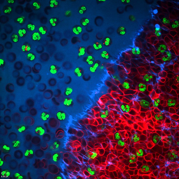
Biopsychronology of a COVID-19 patient blood sample in the ibidi µ-Slide 8 Well after induction of coagulation. The right side shows the area of the blood clot with the fibrin network (blue) and the erythrocytes (red). Note the abundant granulocytes (their nuclei labeled in green) fighting against COVID-19 and other enemies of our health. Image acquisition was performed on a spinning disc confocal microscope.
Published in the 2021 ibidi Calendar.