Stage Top Incubator
Vascular Research
Institute for Molecular Bioscience, The University of Queensland, Brisbane, Australia
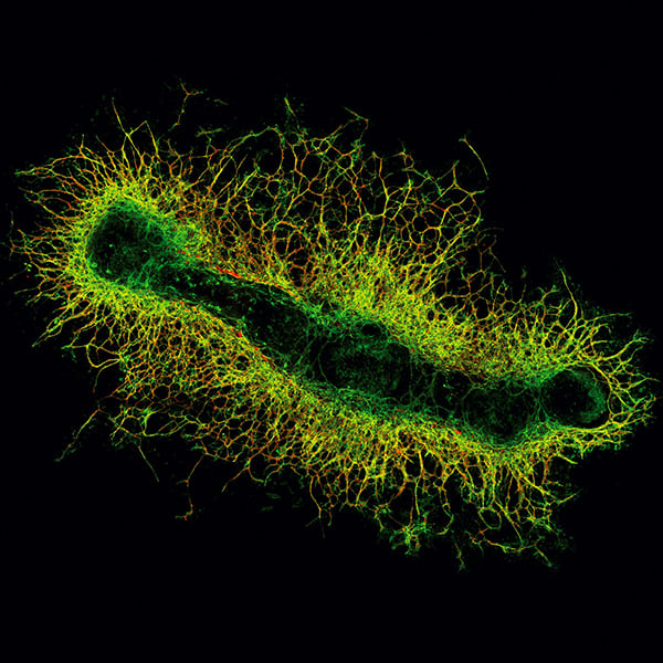
A murine metatarsal as an ex vivo model for sprouting angiogenesis. The fetal mouse bone was obtained from an E16.5 LifeAct-GFP mouse embryo and cultured for 14 days in an ibidi µ-Plate 24 Well, leading to the outgrowth of blood vessels. The metatarsal shows the actin cytoskeleton in green, and was stained for CD31 (PECAM-1) in red as an endothelial marker to highlight vessel outgrowth from the bone.
Published in the 2021 ibidi Calendar.