Stage Top Incubator
Immunology
Universitätsklinikum Hamburg-Eppendorf, Hamburg, Germany
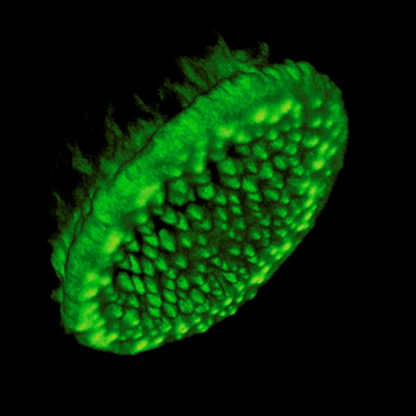
Podosomes in a primary human macrophage. Primary human macrophage isolated from peripheral blood, overexpressing LifeAct-Tag GFP2, highlighting F-actin rich structures. 3D reconstruction from confocal laser scanning micrographs of ventral part of the cell. The F-actin rich dots correspond to the cores of podosomes, adhesion structures of monocytic cells, which are also sites of extracellular matrix degradation. The podosome core has a diameter of approx. 0.3 μm, a height of approx. 0.5 μm, and is localized at the substrate-attached part of the cell.
Published in the 2012 ibidi Calendar.