


More Than Plastic: The ibidi Polymer Coverslip
Why Cell Enthusiasts and Microscopists Equally Love it
ibidi Blog | June 2, 2022 | Tina Freisinger, ibidi GmbH
Have you ever heard the full story about the ibidi Polymer Coverslip? After all, it is the heart of our µ-Slides, µ-Dishes and µ-Plates, and largely contributes to our success.
It all began in the early 2000s when three PhD students and their PI had a vision to found a company that would revolutionize cell-based assays by combining cell culture and high-resolution microscopy. Up until this point, cell biologists had to compromise between surfaces that were either good for cell growth and bad for imaging (e.g., tissue culture flasks that are made of polystyrene) or good for imaging and bad for cell growth (e.g., glass coverslips).
|
|
|
Optical Properties of the ibidi Polymer Coverslip
Scientists who use the ibidi labware with the Polymer Coverslip Bottom for microscopy often don't even realize that it is not made of glass—simply because you can't tell the difference in imaging quality.
So, why is that? There are certain material properties that are essential for high quality microscopy. We will take a closer look at five of these parameters (coverslip thickness, refractive index, Abbe number, transmission, and autofluorescence) and how the specs of the ibidi Polymer Coverslip compare to both glass and conventional polystyrene tissue culture vessels.
Coverslip Thickness
|
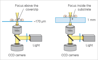
The thickness of the coverslip plays an important role in the imaging quality, because most microscope objective lenses are designed to focus on a specimen on top of a coverslip with a certain thickness (Fig. 1). In cell microscopy, the standard coverslip thickness is 0.17 mm (170 µm +20/–10 µm), which corresponds to a #1.5 coverslip. While most polystyrene tissue culture plates and dishes have a bottom thickness of 500–1000 µm, the ibidi Polymer Coverslip has the same thickness as a standard #1.5 glass coverslip. Refractive IndexThe refractive index (which measures the speed of light inside a specific material) of an ibidi Polymer Coverslip is 1.52, and therefore the same as the refractive index of glass. In contrast, standard tissue culture plates and dishes made from polystyrene have a refractive index of 1.58. This value is important because microscopy objectives are designed for coverslips with a refractive index of 1.52. |
Fig. 1: Microscopic lenses are optimized for the standard coverslip thickness of 170 µm (left). Using other coverslip thicknesses makes it impossible for high resolution objective lenses to focus on the specimen (right). |
Abbe Number
The Abbe number defines the chromatic aberration of optical material. Materials with a higher Abbe number give less color dispersion, and therefore provide a better optical quality for microscopy. A material with an Abbe number greater than or equal to 55 is considered to be well-suited for high-resolution microscopy. The ibidi Polymer Coverslip has an Abbe number of 56, and a standard glass coverslip has an Abbe number of 55. In contrast, polystyrene has an Abbe number of 31.
Transmission
|
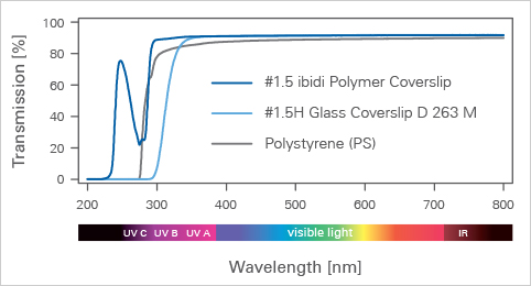
To provide high-quality imaging results, the transmission ability of microscopy carriers has to be high across a variety of different wavelengths. This is the case for glass as well as for the ibidi Polymer Coverslip. Standard polystyrene materials block the UV light, which makes them not really suitable for all microscopy techniques and fluorophores (Fig. 2). |
Fig. 2: Transmission curves of various coverslip and cell culture materials across different wavelengths. |
Autofluorescence
|
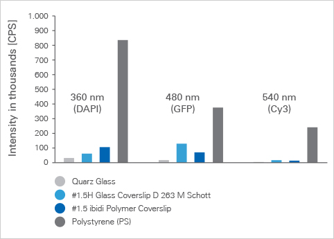
The autofluorescence properties of the microscopy carrier material can disturb the imaging process by showing up as a high background signal during microscopy. While the autofluorescence of the ibidi Polymer Coverslip material and glass is very low, cultureware made from polystyrene has a very high autofluorescence (Fig. 3). |
Fig. 3: Autofluorescence intensity for different coverslip and cell culture materials at different wavelengths. |
Cell Culture Properties of the ibidi Polymer Coverslip
Up to now, we have talked a lot about the optical properties of the ibidi Polymer Coverslip and the reasons why it is so suitable for high-quality imaging, but what about the cells?
Do they feel comfy on this surface? Do they like to attach and grow on it? The answer is simply, YES! And here is why:
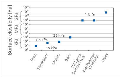
Surface Stiffness
|
First of all, plastic is softer than glass, which makes its surface elasticity more similar to in vivo tissue (Discher et al., 2005; Tissue-Culture TreatmentSecondly, in contrast to glass, the surface of plastic can be modified in a way that makes it more hydrophilic, which most adherent cell types love. This physical surface treatment (which is not a coating) is commonly known as tissue culture treatment. Most plastic vessels used for cell culture are modified in this way. At ibidi, we call the tissue culture surface modification of our Polymer Coverslip, "ibiTreat," which is not a protein coating, but a physical surface treatment that enhances the attachment of the cells. Of course, additional protein coatings can be applied to the uncoated ibidi Polymer Coverslip or the ibiTreat version without compromising the optical quality. |
Fig. 4: Surface elasticity of different tissues as well as coverslip and cell culture materials. |
Gas Permeability
The third reason why the cells like to grow on the ibidi Polymer Coverslip is its gas permeability. This allows gas exchange to take place through the full bottom surface, supplying the cells with fresh O2 and allowing CO2 to leave the cellular environment (Fig. 5).
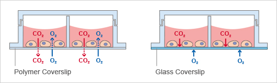
Fig. 5: In contrast to glass (right), the Polymer Coverslip allows for gas exchange across the bottom surface (left).
Given all the above reasons, the ibidi Polymer Coverslip combines the best of two worlds—ideal optics for high-resolution imaging and an optimal surface for cell attachment and growth*.
Still not convinced? Make a side-by-side comparison of the Polymer Coverslip and a Glass Bottom ibidi Imaging Chamber by ordering a free sample today: Free Sample Program
Find out more about the ibidi Imaging Chambers, microscopy parameters of different materials, and ibidi material specifications on our Application Site, "Microscopy with ibidi."
*Disclaimer: There are some applications (e.g., certain coatings or microscopy techniques) where a glass surface is recommended. Find out more on the glass bottom application website.
Reference:
- Discher DE, Janmey P, Wang YL. Tissue cells feel and respond to the stiffness of their substrate. Science. 2005 Nov 18;310(5751):1139-43. doi: 10.1126/science.1116995.
Read article
 (3)
(3)
 (0)
(0)