Stage Top Incubator
Neurobiology
Jozef Stefan Institute, Ljubljana, Slovenia
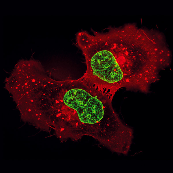
Human microglial cells (HMC3) were stained with Hoechst (nuclei, green) and ATTO 647N DPPE (red), a lipophilic fluorescent probe. After incorporation of the phospholipid into the plasma membrane, the fluorophore is located at the water/lipid interface of the membrane. Over time, it is incorporated into the cell, labeling further structures with lipid membranes. The image was acquired in a µ-Slide 8 Well high Glass Bottom using super-resolution (STED) mounted on an Olympus IX83 microscope with a 60x objective lens.
Published in the 2023 ibidi Calendar.