Stage Top Incubator
Neurobiology
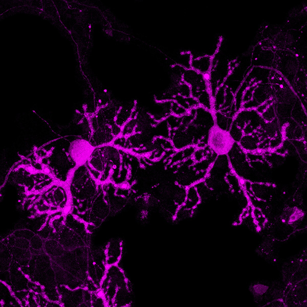
Graduate School of Pharmaceutical Sciences, Nagoya City University, Nagoya, Japan
Fluorescence microscopy image of murine cerebellar Purkinje neurons cultured in an ibidi µ-Dish 35 mm, low Grid-500. The cells were stained against the IP3 receptor (magenta) and imaged using a Zeiss LSM800 confocal microscope with a 40x objective lens.
Published in the 2022 ibidi Calendar.