Stage Top Incubator
Cancer Research
1Health Research Institute Valdecilla (IDIVAL), Spain 2Dept. Anatomy & Cell Biology, University of Cantabria, Santander, Spain
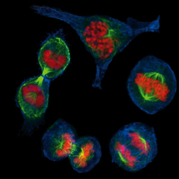
MCF7 breast cancer cells during the different stages of mitosis. Cells were grown on an ibidi Glass Bottom Dish 35 mm and stained with phalloidin (blue) to visualize F-actin, α-Tubulin for the mitotic spindle (green), and DAPI (red) for DNA. Cells were visualized on a NIKON A1R scanning laser confocal microscope using a 60x objective.
Published in the 2021 ibidi Calendar.