




From Petri Dish to Organ-on-Chip
ibidi Blog | September 12, 2026 | Abhishek Derle, ibidi GmbH
How much of human biology can we really capture once cells are taken out of the body?
Conventional cell culture has taught us an enormous amount, and the Petri dish remains one of the most important tools in biology. But cells inside the body experience much more than a surface and culture medium. Blood flows, tissues stretch, nutrients and signaling molecules form gradients, and different cell types constantly communicate with each other. As research questions become more complex, there is growing interest in models that can recreate some of these conditions without losing the control and accessibility of in vitro experiments.
Organ-on-chip technology helps bridge this gap. By combining living cells with microfluidics, defined tissue environments, and controlled physical cues, these systems bring selected aspects of human physiology into the laboratory in a measurable and surprisingly compact way.
So, What Exactly Is an Organ-on-Chip?
Despite the name, an organ-on-chip is not a tiny complete organ sitting inside a plastic device. It is an engineered in vitro model designed to reproduce specific structural, mechanical, biochemical, or functional characteristics of a tissue or organ [1].
These models are often grouped under the broader term microphysiological systems, or MPS. The goal is not to rebuild an entire organ in miniature, but to capture the biological features that matter most for a specific question under controlled experimental conditions. Starting with human cells, researchers can gradually add back the parts of their natural environment that are important for the experiment.

Fig. 1 | From human physiology to organ-on-chip and body-on-chip models. Organ-on-chip systems recreate selected tissue functions, while connected models enable the study of interactions between multiple organs.
Flow can recreate mechanical forces, additional cell types can introduce tissue interactions, separate compartments can mimic barriers or communication between neighboring tissues, and 3D matrices can provide a more tissue-like architecture. In other words, the cells get a little more of their biological context back.
This flexibility is one of the major strengths of organ-on-chip technology. A model can stay focused on a single tissue, such as a vessel, lung, gut, or brain, and reproduce only the functions needed for the experiment. But it does not have to stop there. Several organ models can also be connected to create multi-organ or body-on-chip systems, allowing researchers to investigate how different tissues communicate and influence one another. The model can therefore grow with the biological question: from isolated human cells, to one organ model, and finally to a connected system when interactions between organs become part of the story.
Microfluidics: Giving Cell Culture a Circulation
One of the key technologies behind many organ-on-chip systems is microfluidics, the controlled movement of very small liquid volumes through precisely defined channels. At first glance, this may sound like miniature laboratory plumbing. Biologically, however, controlled flow can completely change the environment surrounding the cells [2]. In blood vessels, for example, flowing liquid generates wall shear stress. Endothelial cells sense these forces and respond by changing their morphology, alignment, signaling, and inflammatory state, meaning that different shear conditions and matrix properties can lead to very different endothelial responses [3].
Perfusion also keeps things moving in other ways: nutrients and oxygen can be continuously supplied, metabolic waste removed, signaling molecules transported, and chemical gradients generated. And because flow rate and channel geometry can be adjusted, researchers can tune the mechanical environment to the needs of the biological model rather than relying on one standard condition. This makes choosing the right flow setup for the experiment an important part of the design. Microfluidics can also bring different cell populations into controlled contact: tumor cells can invade through a matrix, immune cells can travel over an endothelial layer, and neighboring tissues can exchange soluble signals across defined compartments.
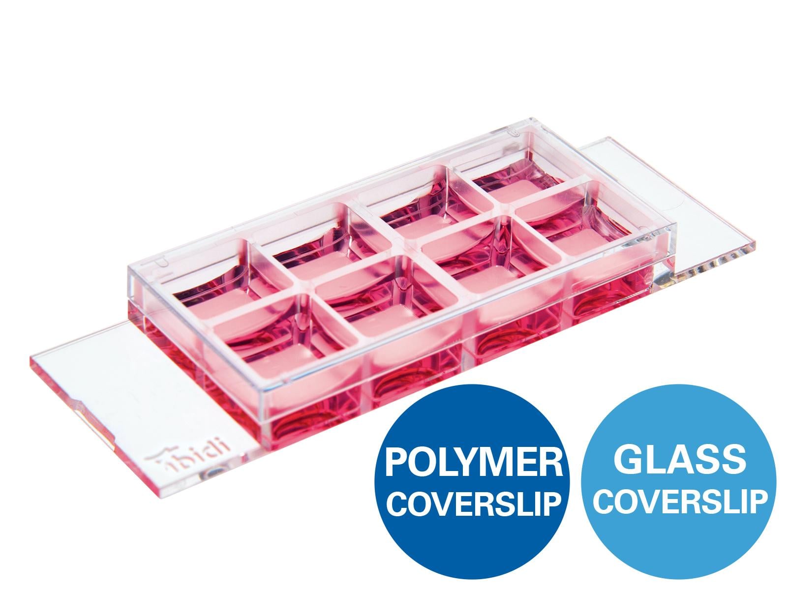
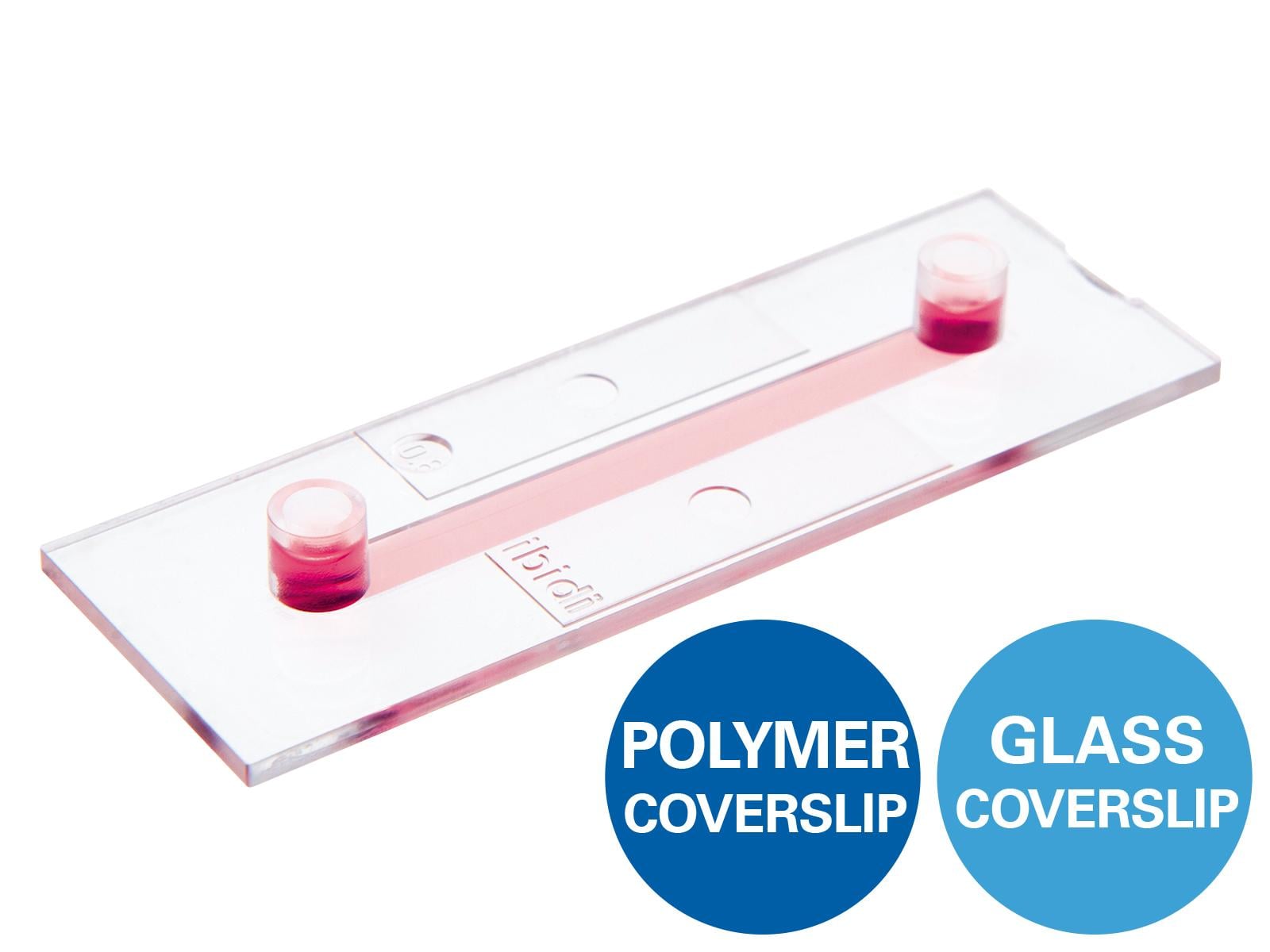
Different microfluidic formats can then add the level of complexity the model needs. The ibidi µ-Slide I Luer 3D, for example, enables cells to be cultured on a 3D gel matrix under defined flow, while the µ-Slide Tissue Engineering provides more freedom to create customized 3D microenvironments and perfusable tissue models. Combined with the ibidi Pump System, these setups allow controlled long-term perfusion and different flow profiles. And once the biology starts responding, the microscopy-compatible slide bottoms keep those changes directly accessible for live cell and high-resolution imaging.
One Principle, Many Organs
The engineering may be similar, but the biology can look completely different from one chip to the next. Brain-on-chip models may combine neurons, glial cells, extracellular matrices, and vascular components to reproduce aspects of neuronal circuits, neuroinflammation, or the blood-brain barrier [4]. Kidney-on-chip models can recreate features such as filtration barriers, tubular structures, and fluid flow, providing a controlled way to investigate renal function and tissue responses in vitro [2].
Gut-on-chip systems take this idea in yet another direction. They can combine an epithelial barrier with fluid flow, peristalsis-like mechanical movements, immune components, and even living microbial communities, bringing several features of the intestinal environment together in one experimental model [5]. Engineered mini-colons take this concept a step further by combining organoid biology with a defined tissue architecture that supports long-term culture, continuous epithelial renewal, and more in vivo-like cellular diversity and organization [6]. Rather than simply keeping the tissue alive, the model maintains an active balance between cell production and cell shedding, capturing an important feature of how the intestinal epithelium normally renews itself. This makes the system especially interesting for studying tissue homeostasis over longer periods under controlled in vitro conditions.
What changes from model to model is not the basic principle, but the biology researchers choose to bring into focus.

Fig. 2 | Long-term cell renewal in bioengineered human mini-colons. The GIF follows the mini-colon epithelium as it grows and continuously renews over time. Shed cells are removed from the lumen, helping maintain a stable tissue structure and long-term homeostasis. From Mitrofanova et al. (2024).
Beyond One Organ: Connected and Vascularized Systems
Human organs do not work in isolation, and increasingly, neither do organ-on-chip models. Connecting different tissues allows researchers to study how signals, metabolites, and drugs travel from one organ model to another. Cardiac microtissues and kidney organoids, for example, have been cultured in separate but communicating chambers of the ibidi µ-Slide III 3D Perfusion and maintained under unidirectional flow with the ibidi Pump System. This created a cardiorenal model in which both tissues retained organ-specific structural and functional features while sharing the same circulating environment [7]. That kind of setup gets closer to an important reality of human physiology: what happens in one tissue rarely stays there.

Fig. 3 | Connecting cardiac and kidney tissues under controlled flow. Cardiac microtissues (cMTs) and kidney organoids (kOs) were placed in separate but communicating chambers of the ibidi µ-Slide III 3D Perfusion and cultured under flow with the ibidi Pump System for 72 hours. The setup was designed to study heart–kidney communication while keeping both tissue models physically separated and functionally accessible for downstream analysis. From Gabbin et al. (2023).
Vascularization adds another layer. As 3D tissues become larger and more complex, diffusion alone is not enough to supply every cell efficiently. Perfusable vascular networks can improve transport while creating a more realistic interface between tissue and circulation. In a 2024 Nature Communications study, endothelial networks formed around spheroids and vascular organoids and were connected to functional intravascular perfusion, showing how microfluidics can bring vascular supply directly into complex 3D models [8].
From a Sketch to a Microenvironment
Not every organ-on-chip model needs to begin with a fixed channel geometry. Researchers can also build tissue-specific microenvironments using hydrogels and light-based microfabrication. Photocrosslinkable materials can be structured into channels, barriers, compartments, or scaffolds, allowing the physical architecture of the experiment itself to become a controllable variable [2].
A narrow endothelialized channel may reproduce aspects of a small blood vessel, while a hydrogel compartment can support 3D invasion or vascular network formation. The ibidi Micro Illumination System enables these geometries to be created using 365 nm UV illumination and customized photomasks, making it possible to generate and adapt defined hydrogel structures for perfusable microfluidic models. This way, geometry can be designed around the biology, rather than forcing the biology into a predefined shape.

Fig. 4 | Light-induced microfabrication for customized microfluidic models. The ibidi Micro Illumination System enables defined patterning of photocrosslinkable hydrogels using UV illumination and customized photomasks to create tailored, perfusable microfluidic models.
Conclusion: Adding the Right Context
Organ-on-chip technology is not about replacing every Petri dish with a microfluidic device. It is about recognizing when cells need more of their physiological environment to answer the question at hand. Flow, gradients, tissue interfaces, mechanical forces, vascularization, and communication between tissues can now be introduced under controlled in vitro conditions.
As these models become more sophisticated, the challenge is to keep them reproducible, scalable, and easy to compare between laboratories. Standardization and clearly defined functional readouts will therefore be increasingly important [9]. The goal is not to build the most complicated chip possible, but the right model to capture the biology that matters.
References
- Vunjak-Novakovic, G., K. Ronaldson-Bouchard, and M. Radisic, Organs-on-a-chip models for biological research. Cell, 2021. 184(18): p. 4597-4611.
- Leung, C.M., et al., A guide to the organ-on-a-chip. Nature Reviews Methods Primers, 2022. 2(1): p. 33.
- Walther, B.K., et al., Mechanotransduction-on-chip: vessel-chip model of endothelial YAP mechanobiology reveals matrix stiffness impedes shear response. Lab Chip, 2021. 21(9): p. 1738-1751.
- Servais, B., et al., Engineering brain-on-a-chip platforms. Nature Reviews Bioengineering, 2024. 2(8): p. 691-709.
- Özkan, A., et al., Intestinal organ chips for disease modelling and personalized medicine. Nature Reviews Gastroenterology & Hepatology, 2024. 21(11): p. 751-773.
- Mitrofanova, O., et al., Bioengineered human colon organoids with in vivo-like cellular complexity and function. Cell Stem Cell, 2024. 31(8): p. 1175-1186.e7.
- Gabbin, B., et al., Heart and kidney organoids maintain organ-specific function in a microfluidic system. Materials Today Bio, 2023. 23: p. 100818.
- Quintard, C., et al., A microfluidic platform integrating functional vascularized organoids-on-chip. Nature Communications, 2024. 15(1): p. 1452.
- Nahon, D.M., et al., Standardizing designed and emergent quantitative features in microphysiological systems. Nature Biomedical Engineering, 2024. 8(8): p. 941-962.

Article written by Abhishek Derle, PhD
ibidi GmbH | September 12, 2026
Biomedical scientist with expertise in cancer biology, metabolism research, and advanced microscopy. Abhishek received his PhD in Biomedical Sciences and Oncology, with research focusing on intracellular trafficking, metabolic regulation, and cellular disease mechanisms.
 (0)
(0)  (0)
(0)