



Cell Co-Culture Assays and Models
Cell co-culture is the cultivation of two or more cell populations within one experimental system so that their communication or interaction can be studied. Depending on the setup, the populations may exchange soluble factors, establish direct cell–cell contact, interact across a porous membrane, or meet at a hydrogel interface.
A co-culture assay is useful when the experimental readout depends on interactions between cell populations, such as paracrine signaling, immune synapse formation, tumor–stroma interaction, barrier function, transmigration, or cell-derived chemotaxis. A monoculture is generally the clearer choice when the aim is to isolate a cell-autonomous response without signals from another population.
ibidi provides specialized systems for direct 2D and 3D co-culture, indirect 2D and 3D co-culture through shared medium, membrane-based and hydrogel-interface models, dynamic co-culture under flow, and cell-generated chemotactic gradients. The guide below gives an overview of relevant in vitro models.
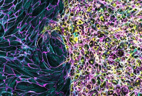
Endothelial cells (left) and trophoblast cells (right) were cultured in an ibidi µ-Dish 35 mm, high with a Culture-Insert 2 Well. Cells were allowed to migrate towards each other after removal of the insert prior to fixation, staining, and imaging. Cells were stained with an antibody against VE-cadherin (magenta), phalloidin for F-actin (cyan), and Cytokeratin 8 (yellow). Image used with permission from Derek Sung.
Which Co-Culture Model Should I Use?
Start with the biological interaction that must be preserved. Then decide whether the cell populations need to touch, remain experimentally separable, share only soluble factors, interact with an extracellular matrix, cross a barrier, generate a chemotactic signal, or experience controlled perfusion. The imaging method, required throughput, culture medium, cell ratio, and order of seeding should be considered before selecting the labware.
| Co-Culture Model | When to Use It | Experimental Setup |
|---|---|---|
| Direct 2D Co-Culture | Use when different 2D cell populations should establish physical contact, either after spatially defined seeding or from the beginning of the assay. | Populations are seeded separately and allowed to establish contact, positioned on defined adhesive areas, or mixed in the same vessel from the start. |
| Direct 3D Co-Culture | Use when different cell types should establish direct contact within or on a shared 3D matrix. | Cell types are mixed within the same hydrogel or extracellular matrix, incorporated into the same spheroid or organoid, or combined on and within a gel. |
| Indirect 2D and 3D Co-Culture Through Shared Medium | Use when 2D or 3D populations should exchange soluble factors without mixing, with or without an extracellular matrix. | Cell populations are cultured in separate wells connected by shared medium, either on 2D surfaces or embedded within or seeded on a 3D extracellular matrix. |
| Membrane-Based 2D and 3D Co-Culture | Use when populations should remain separated by a porous membrane while soluble factors or cells can pass through its pores. | Cell layers are cultured on one or both membrane surfaces, optionally with a second population embedded in a 3D matrix in an adjacent compartment. |
| 3D Co-Culture Across Hydrogel Interfaces | Use when populations should remain spatially organized in, on, or next to a 3D hydrogel without a porous membrane. | Populations are positioned in adjacent gel compartments or on and within the same gel without initially mixing them. |
| Dynamic 2D and 3D Co-Culture Under Controlled Flow | Use when perfusion, wall shear stress, or flow-dependent signaling is part of the biological question. | In 2D models, adherent cell layers interact with cells in the flowing medium. In 3D models, cells are cultured in or on a gel under controlled perfusion. |
| 2D and 3D Co-Culture With Cell-Generated Chemotactic Gradients | Use when one living cell population should generate the chemotactic signal that guides another population. | Signal-producing cells are seeded in one reservoir, while migrating cells are cultured in the central observation area on a 2D surface or within a 3D gel. |
Direct 2D Co-Culture
Direct 2D co-culture can begin with spatially separated populations or with different cell types mixed in the same culture vessel. The appropriate setup depends on whether the assay should capture migration and first contact or interactions between cells that are mixed from the start.
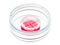
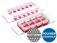
The ibidi Culture-Inserts keep adherent populations separated during attachment. After insert removal, a defined cell-free gap allows the populations to migrate toward each other and establish contact. Micropatterned Labware provides an alternative when cells should be positioned on defined adhesive areas before another population is added.
For mixed 2D co-culture without physical divisions, Chambered Coverslips, Dishes, and Multiwell Plates provide microscopy-compatible formats with different culture areas and throughput levels. These formats can be used, for example, for tumor–immune co-culture, immune synapse imaging, and cell killing assays.
Detailed method guidance is available on the Wound Healing and Migration application page and the Micropatterning application page.
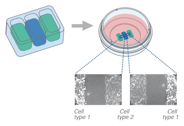
Spatially separated cell populations can be seeded in the individual wells of an ibidi Culture-Insert. After insert removal, the cell fronts can migrate toward each other and interact.
ibidi Solutions for Direct 2D Co-Culture
Start with Separated Cell Populations:
Start with Mixed Cell Populations:

Direct 3D Co-Culture
Direct 3D co-culture combines different cell types within or on a shared extracellular matrix, hydrogel, or scaffold, or within multicellular structures such as spheroids and organoids. Because the populations occupy the same 3D environment, they can establish direct cell–cell contact while also interacting through soluble factors and the surrounding matrix.
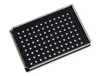
The micro-Insert 3D, µ-Slide 15 Well 3D, and µ-Plate 96 Well 3D support configurations in which different populations are mixed within a gel or combined on and within the same matrix.
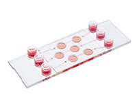
The µ-Slide I Luer 3D and µ-Slide III 3D Perfusion extend these configurations to channel-based models and can also be used with controlled perfusion when flow is part of the experimental design.
For general guidance on matrices, spheroids, organoids, and assay development, see the 3D Cell Culture application page.
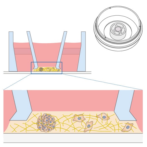
Different cell types share the same extracellular matrix in the micro-Insert 3D.
ibidi Solutions for Direct 3D Co-Culture
Indirect 2D and 3D Co-Culture Through Shared Medium
Indirect 2D and 3D co-culture through shared medium keeps cell populations physically separated while allowing soluble factors to move between them. The populations can be cultured with or without an extracellular matrix, making the setup suitable for paracrine signaling and feeder-cell effects when direct cell–cell contact is not required.
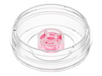
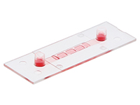
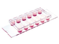
The µ-Slide 2 Well Co-Culture and the micro-Insert 4 Well allow each population to be seeded and cultured in a separate well. In 2D setups, cells grow on the coverslip bottom. In matrix-based 3D setups, one or both populations can be embedded within or seeded on an extracellular matrix. The wells are then connected by shared medium, enabling soluble communication while the populations remain spatially separated and individually accessible.
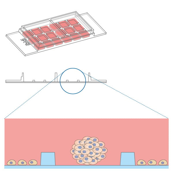
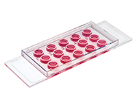
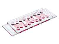
Different cell populations can be spatially organized in the µ-Slide 2 Well Co-Culture while communicating through shared medium.
ibidi Solutions for Indirect 2D and 3D Co-Culture Through Shared Medium
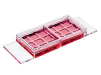
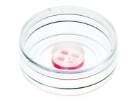
Application Note
Membrane-Based 2D and 3D Co-Culture
Membrane-based co-culture keeps populations on opposite sides of a porous barrier while soluble molecules pass between the compartments. Depending on the pore size, membrane properties, and assay design, cells may also migrate through the membrane.
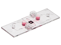
The µ-Slide ibiPore SiN supports the cultivation of cell layers on one or both sides of a porous silicon nitride membrane. It can also combine a cell layer on the membrane with a second population embedded in a 3D gel.
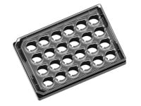
The µ-Plate 24 Well for Membrane Inserts provides microscopy access for assays using compatible third-party porous membrane inserts. The membrane inserts are separate products and are not included with the plate.
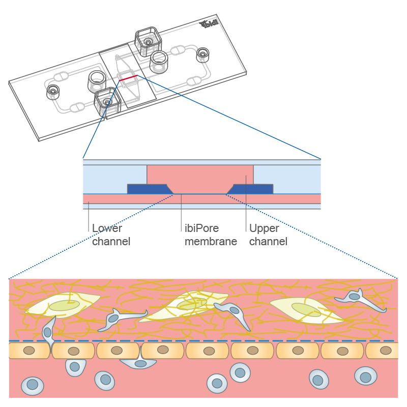
A membrane-based co-culture can combine a cell layer on the µ-Slide ibiPore SiN membrane with a second population embedded in a 3D gel.
ibidi Solutions for Membrane-Based 2D and 3D Co-Culture
3D Co-Culture Across Hydrogel Interfaces
3D co-culture across hydrogel interfaces keeps populations in defined positions within, on, or next to a gel without a porous membrane. In contrast to direct 3D co-culture with initially mixed populations, the cell types begin in separate regions and interact across the matrix interface.
The micro-Insert 3D uses a central hydrogel to create inner and outer compartments that remain accessible from both sides. This membrane-free geometry supports configurations with cells within the gel, on the gel surface, or in the surrounding compartment.
The µ-Slide I Luer 3D, µ-Slide 15 Well 3D, and µ-Plate 96 Well 3D support additional arrangements in which different populations are positioned on and within a hydrogel. The choice depends on whether the experiment requires a channel format, microscopy access, or higher throughput.
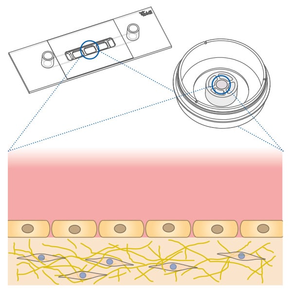
3D co-culture with defined cell positions: one cell population is located in the gel and a second cell population on the gel surface using the micro-Insert 3D or the µ-Slide I Luer 3D.
ibidi Solutions for 3D Co-Culture Across Hydrogel Interfaces
Dynamic 2D and 3D Co-Culture Under Controlled Flow
Dynamic co-culture under controlled flow is appropriate when perfusion, wall shear stress, or flow-dependent signaling is part of the biological question. In rolling and adhesion assays, one cell type, such as endothelial cells, is cultured in the channel while a second population, such as leukocytes, is introduced with the flowing medium.
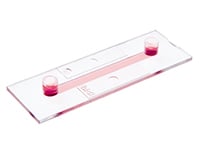
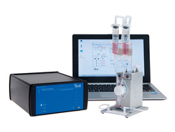
The µ-Slide I Luer and µ-Slide VI support 2D flow-based co-culture. The µ-Slide I Luer 3D and µ-Slide III 3D Perfusion extend controlled flow to 3D configurations with cells or spheroids in or on a gel. The ibidi Pump System provides controlled long-term perfusion.
Detailed design of dynamic 3D models and perfusion is covered on the advanced 3D culture and perfusion application page.
For flow profiles, pump setup, and wall shear stress, see the Cell Culture Under Flow application page.
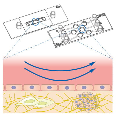
A dynamic 3D co-culture can combine cells or spheroids in and on a gel matrix with controlled medium flow.
ibidi Solutions for Dynamic 2D and 3D Co-Culture Under Controlled Flow
2D and 3D Co-Culture With Cell-Generated Chemotactic Gradients
Cell-generated chemotactic gradients provide a specialized indirect co-culture setup in which one living cell population produces the migration signal and the directed response of another population is measured.
The µ-Slide Chemotaxis can be used when signal-producing cells are cultured in one reservoir and migrating cells are placed in the central observation area. Depending on the validated protocol, the migrating population can be analyzed on a 2D surface or within a 3D gel.
Experiments using conditioned medium or purified chemoattractants are chemotaxis assays, but they are not co-cultures within the slide.
For gradient generation and quantitative migration analysis, see the Chemotaxis Application Guide.
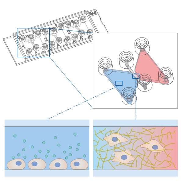
Cells in one reservoir of the µ-Slide Chemotaxis produce a chemoattractant that forms a stable gradient across the observation area, enabling co-culture chemotaxis assays in 2D or 3D.
ibidi Solution for Cell-Generated Chemotactic Gradients
Frequently Asked Questions About Cell Co-Culture
What Is a Cell Co-Culture Assay?
A cell co-culture assay combines two or more distinct cell populations in one experimental system to study cell–cell communication or interaction. Depending on the design, the populations can establish direct contact or communicate while separated by shared medium, a porous membrane, or a hydrogel interface.
What Is the Difference Between Co-Culture and Monoculture?
A monoculture contains one cell population and is appropriate for studying cell-autonomous responses. A co-culture contains two or more populations and is used when paracrine signaling, direct cell–cell contact, matrix interaction, barrier behavior, immune-cell interaction, or another heterotypic response is part of the biological question.
What Is the Difference Between Direct and Indirect Co-Culture?
In direct 2D co-culture or direct 3D co-culture, different cell populations can establish physical contact. In indirect co-culture, they remain spatially separated but exchange soluble factors through shared medium, a porous membrane, or a hydrogel interface. Culture-Inserts enable separate seeding followed by direct interaction after insert removal, while the µ-Slide 2 Well Co-Culture allows populations to share medium without mixing.
What Is the Difference Between 2D and 3D Co-Culture?
In 2D co-culture, cells grow mainly as monolayers on a flat culture surface. In 3D co-culture, different populations interact within or on an extracellular matrix or hydrogel, or within multicellular structures such as spheroids and organoids. The appropriate format depends on whether matrix-dependent behavior and three-dimensional spatial organization are relevant to the experimental question. General model development is covered on the 3D Cell Culture application page.
How Do I Choose a Co-Culture Setup?
First decide whether direct contact is required and whether the populations must remain experimentally separable. Then select a 2D or 3D format and determine whether the model needs shared medium, a porous membrane, a hydrogel interface, a cell-generated chemotactic signal, or controlled flow. Finally, match the geometry and throughput to the imaging method and experimental readout.
How Can Cell Populations Be Kept Separate in an Indirect Co-Culture?
Cell populations can be cultured in separate wells connected by shared medium, on opposite sides of a porous membrane, or in defined regions separated by a hydrogel interface. The appropriate architecture depends on whether the experiment requires soluble-factor exchange alone, cell passage, barrier function, cell–matrix interaction, or separate access to each population.
Which Parameters Should Be Optimized in a Co-Culture Assay?
Important variables include culture medium compatibility, cell ratio, seeding density, seeding order and timing, matrix composition when applicable, assay duration, and the imaging or endpoint readout. Monoculture controls should be included for each population. Depending on the biological question, conditioned medium or contact-blocking controls can help distinguish soluble-factor effects from contact-dependent interactions.