


Cell Culture Under Flow: Applications and Experimental Examples
This page summarizes common cell culture under flow applications and wall shear stress assay applications for cell models exposed to defined fluid flow under controlled in vitro conditions. It is intended for researchers planning endothelial, epithelial, vascular, immune cell interaction, barrier, mechanotransduction, and 3D disease model studies under flow.
The focus is on selecting the right application for your biological question. Depending on the model, cells can be cultured as 2D monolayers under flow, in matrix-based 3D models, or in membrane-based co-cultures using optically suitable porous membranes. Each application chapter connects the biological context with experimental examples and suitable ibidi solutions.
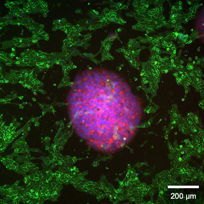
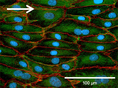
Staining of spheroids (blue, red) and HUVECs (green) after 3 days in co-culture under perfusion. Cell nuclei: DAPI (blue); actin filaments: Phalloidin-iFluor 647 (red); CD31: Alexa Fluor 488 labeled anti-CD31 antibody (green).
In brief: Wall shear stress assays are used to study how adherent cells respond to defined fluid flow. Major applications include endothelial cell conditioning, disturbed-flow models, barrier models, rolling and adhesion assays, and cell interaction studies.
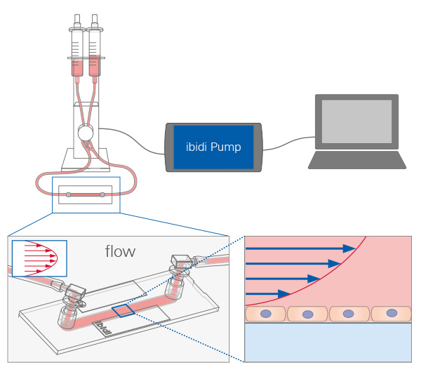
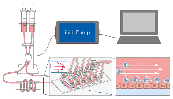
For learning the biological background of flow assays, see the Wall Shear Stress and Flow Types chapter. To plan and conduct a flow assay in your lab, see the Experimental Workflow page.
Find the Right Flow Application for Your Research
Cell culture under flow can be used for a wide range of applications, such as:
- Endothelial mechanobiology, shear stress response, and vascular flow conditioning
- Lymphatic endothelial flow biology
- Atherosclerosis, disturbed flow, and vascular dysfuncion
- Thrombosis and platelet adhesion under flow
- Immune cell recruitment, interaction, and transendothelial migration
- Barrier models, permeability, and blood–brain barrier research
- Kidney tubule epithelial models and nephrotoxicity
- Airway epithelial flow and mucociliary biology
- Gut epithelial barrier and host–microbiome interaction
- Tumor microenvironment, extravasation, and drug delivery
- Viral and bacterial infection under flow
Many additional cell culture under flow applications are possible depending on the cell type, flow profile, culture format, and readout. If your specific research question is not listed here, our application specialists can help you choose the most suitable ibidi flow setup. Please contact us for individual support.
The table below highlights selected high-priority application areas that can be addressed with ibidi flow products and are described in more detail in the following chapters.
| Research Application | Suggested Cell Culture Model | ibidi Solutions |
|---|---|---|
| Endothelial Mechanobiology and Vascular Flow Conditioning | 2D endothelial monolayer under defined laminar flow | ibidi Pump System, µ-Slide I Luer, µ-Slide VI |
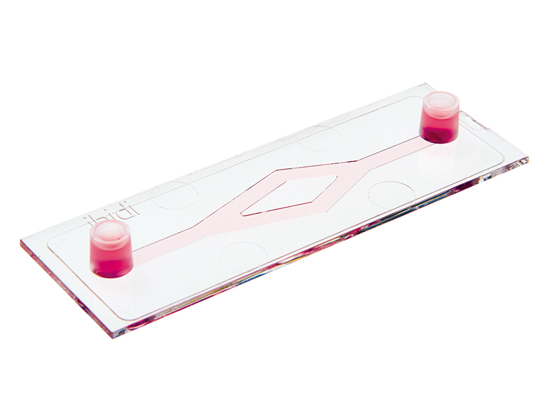
| Atherosclerosis, Disturbed Flow, and Vascular Dysfunction | 2D endothelial monolayer under laminar, disturbed, or branched flow | ibidi Pump System, µ-Slide y-shaped, µ-Slide I Luer |
| Immune Cell Recruitment, Interaction, and Transendothelial Migration | Activated endothelial monolayer under flow combined with suspended immune cells | ibidi Pump System, µ-Slide VI, µ-Slide I Luer, µ-Slide I Luer 3D |
| Barrier Models and Permeability | Endothelial or epithelial barrier model under controlled flow, with or without gel matrix | ibidi Pump System, µ-Slide ibiPore SiN, µ-Slide I Luer |
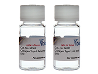
| Tumor Microenvironment, Extravasation, and Drug Delivery | Matrix-based 3D model (spheroids, tumor cells), or co-culture system under flow | ibidi Pump System, µ-Slide I Luer 3D, µ-Slide III 3D Perfusion, Collagen Type I |
Endothelial Mechanobiology and Vascular Flow Conditioning
Endothelial cells are highly sensitive to wall shear stress. Defined laminar flow can be used to condition endothelial monolayers and to analyze morphology, alignment, cytoskeletal organization, junction formation, inflammatory signaling, gene expression, and mechanotransduction.
Culturing Human Umbilical Vein Endothelial Cells (HUVECs) Under Flow
Immunofluorescence staining allows the comparison of static and flow-cultured endothelial cells and can reveal differences in cytoskeletal organization. In this example, flow-conditioned HUVECs were stained against VE-cadherin and compared with static cultures.
VE-cadherin staining highlights adherens junctions in both static and flow-conditioned HUVECs. Under static conditions, the cells are generally larger and show a less organized actin cytoskeleton. In contrast, flow-conditioned cells are oriented and display distinct F-actin stress fibers, indicating flow-dependent reorganization of the endothelial cell layer.
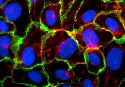
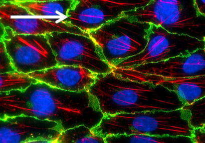
HUVECs were cultured for 5 days under static conditions (left, 0 dyn/cm2) or under flow at 10 dyn/cm2 (right) in a µ-Slide I 0.4 Luer ibiTreat, using the ibidi Pump System. The cells were stained for VE-cadherin (green), F-actin (red), and nuclei (blue). Imaging was performed using a Nikon Eclipse microscope at 60× magnification.
Culturing Pulmonary Endothelial Cells (HPMECs) Under Shear Stress
When cultured under flow, human pulmonary microvascular endothelial cells (HPMECs) show changes in morphology, cytoskeletal organization, and endothelial cell–cell contacts compared with static culture. This example demonstrates how phase contrast and fluorescence microscopy can be used to assess endothelial responses to defined shear stress conditions.

Fluorescence microscopy of HPMECs comparing static culture (left) and flow culture (right) after 72 h in the µ-Slide I 0.6 Luer, coated with Collagen IV. A flow of 2.3 dyn/cm2 was applied using the ibidi Pump System. Fluorescence labels: β-actin (green), VE-cadherin (red), and DAPI (blue). Scale bars 100 µm. Data by Daniel Bourquain, Robert Koch Institute, Berlin.
ibidi Solutions for Endothelial Mechanobiology and Vascular Flow Conditioning
Selected Publications for Endothelial Mechanobiology and Vascular Flow Conditioning
Human arterial endothelial cells were cultured in the µ-Slide I 0.4 Luer and µ-Slide y-shaped and exposed to flow using the ibidi Pump System to study mechanosensitive calcium signaling and anti-inflammatory responses.
Hong SG, Ashby JW, Kennelly JP, Wu M, Steel M, Chattopadhyay E, Foreman R, Tontonoz P, Tarling EJ, Turowski P, Gallagher-Jones M, Mack JJ. (2024) Mechanosensitive membrane domains regulate calcium entry in arterial endothelial cells to protect against inflammation. J Clin Invest. 134(13):e175057, 10.1172/JCI175057.
Read article
Human arterial endothelial cells were cultured in the µ-Slide I 0.4 Luer and exposed to shear stress using the ibidi Pump System to analyze shear stress-modulated chromatin accessibility.
Jatzlau J, Mendez PL, Altay A, et al. Fluid shear stress-modulated chromatin accessibility reveals the mechano-dependency of endothelial SMAD1/5-mediated gene transcription. iScience. 2023;26(9):107405. doi:10.1016/j.isci.2023.107405.
Read article
Vascular endothelial cells were cultured in the µ-Slide I 0.6 Luer and exposed to shear stress using the ibidi Pump System to study flow-dependent regulation of mitochondrial Ca2+ uniporter complex subunit expression.
Patel A, Pietromicca JG, Venkatesan M, et al. Modulation of the mitochondrial Ca2+ uniporter complex subunit expression by different shear stress patterns in vascular endothelial cells. Physiol Rep. 2023;11(3):e15588. doi:10.14814/phy2.15588.
Read article
Atherosclerosis, Disturbed Flow, and Vascular Dysfuncion
Atherosclerosis-related mechanisms are strongly linked to local flow patterns. Non-uniform, oscillatory, or disturbed-flow-like conditions can be used to analyze endothelial activation, inflammatory signaling, monocyte adhesion, mechanosensitive membrane domains, and vascular dysfunction.
Disturbed Flow in a Custom In Vitro Arteriovenous Fistula Model
Disturbed flow plays an important role in endothelial dysfunction and vascular remodeling in arteriovenous fistulas. In this study by Xiao et al., the authors established a custom in vitro arteriovenous fistula (AVF) model that formed a 60° bifurcated flow system. Human umbilical vein endothelial cells were seeded on a transparent polycarbonate membrane (PCM), which was inserted into the model.
To better mimic AVF-like pulsatile hemodynamics, the authors connected the model to a modified ibidi Pump System. A side tube with a small-diameter needle was added upstream of the main tube, allowing residual low-flow perfusion during the low-flow phase instead of complete flow interruption. By adjusting the pump pressure, the setup generated alternating high- and low-flow phases. This allowed the authors to correlate computational fluid dynamics with endothelial morphology, focal adhesion junctions, glycocalyx organization, and flow-sensitive marker expression in defined regions of laminar, oscillatory, and disturbed flow.
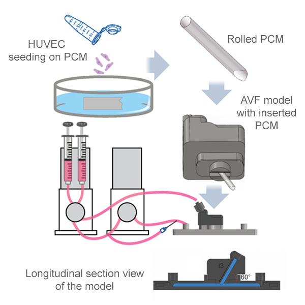
Schematic representation of a custom in vitro arteriovenous fistula model for studying disturbed flow and endothelial cell function. A tailored, cell-seeded transparent polycarbonate membrane was inserted into a 60° bifurcated flow model and perfused using a modified ibidi Pump System to generate pulsatile AVF-like flow conditions. Adapted from: Xiao Z, et al. (2026), doi:10.1016/j.actbio.2026.01.044, licensed under CC BY 4.0.
ibidi Solutions for Atherosclerosis, Disturbed Flow, and Vascular Dysfunction
Selected Publications for Atherosclerosis, Disturbed Flow, and Vascular Dysfunction
HUVECs were cultured on a tailored polycarbonate membrane in a custom-made in vitro arteriovenous fistula model and perfused using the ibidi Pump System to simulate disturbed flow conditions.
Xiao Z, White NA, Wen J, Postma RJ, Sol WMPJ, van den Berg BM, van Zonneveld AJ, van de Stadt HJF, Mirza A, Bijkerk R, Rotmans JI. (2026) Exploring the link between disturbed flow and endothelial cell function in an in vitro arteriovenous fistula model. Acta Biomater., 10.1016/j.actbio.2026.01.044.
Read article
Human aortic endothelial cells were cultured in µ-Slide I Luer variants and exposed to laminar or disturbed flow using the ibidi Pump System to analyze flow-dependent changes in the endothelial lipidome and transcriptome.
Hong SG, Kennelly JP, Williams KJ, Bensinger SJ, Mack JJ. (2024) Flow-mediated modulation of the endothelial cell lipidome. Front Physiol. 15:1431847, 10.3389/fphys.2024.1431847.
Read article
Human coronary artery endothelial cells were cultured in µ-Slide I 0.4 Luer and conditioned with shear stress using the ibidi Pump System to study neutrophil microvesicle and monocyte adhesion to atheroprone endothelium.
Gomez I, Ward B, Souilhol C, Recarti C, Ariaans M, Johnston J, Burnett A, Mahmoud M, Luong LA, West L, Long M, Parry S, Woods R, Hulston C, Benedikter B, Niespolo C, Bazaz R, Francis S, Kiss-Toth E, van Zandvoort M, Schober A, Hellewell P, Evans PC, Ridger V. (2020) Neutrophil microvesicles drive atherosclerosis by delivering miR-155 to atheroprone endothelium. Nat Commun. 11:214, 10.1038/s41467-019-14043-y.
Read article
Human lymphatic endothelial cells were cultured on fibronectin-coated µ-Slide I 0.8 Luer and exposed to oscillatory or laminar flow using the ibidi Pump System to study FOXC2-dependent responses to disturbed flow conditions.
Sabine A, Bovay E, Demir CS, Kimura W, Jaquet M, Agalarov Y, Zangger N, Scallan JP, Graber W, Gulpinar E, Kwak BR, Mäkinen T, Martinez-Corral I, Ortega S, Delorenzi M, Kiefer F, Davis MJ, Djonov V, Miura N, Petrova TV. (2015) FOXC2 and fluid shear stress stabilize postnatal lymphatic vasculature. J Clin Invest. 125(10):3861–3877, 10.1172/JCI80454.
Read article
Immune Cell Recruitment, Interaction, and Transendothelial Migration
Flow assays are widely used to study immune cell and leukocyte rolling, adhesion, and transmigration. In these assays, immune cells are perfused over an activated endothelial monolayer, a protein-coated surface, a membrane, or a matrix-based model. Readouts can include rolling velocity, adhesion count, transmigration events, and live cell tracking.
Rolling and Adhesion Assay Under Defined Shear Stress
Rolling and adhesion assays are commonly used to analyze leukocyte–endothelial interactions under flow. In this example, cerebrovascular endothelial cells (CVECs) were cultured as a 2D endothelial monolayer in the µ-Slide VI 0.4 and stimulated with LPS to induce an inflammatory endothelial phenotype. Granulocytes were then perfused over the activated endothelial layer using the ibidi Pump System at 1 dyn/cm2, enabling live microscopic analysis of granulocyte rolling, adhesion, and cell–cell interactions under defined shear stress.
Rolling and adhesion assay with granulocytes perfused over an LPS-stimulated cerebrovascular endothelial cell monolayer in the µ-Slide VI 0.4. The assay enables visualization of leukocyte–endothelial interactions under defined shear stress. Image courtesy of Gediminas Cepinskas, University of Western Ontario, Canada.
ibidi Solutions for Immune Cell Recruitment, Interaction, and Transendothelial Migration
Selected Publications for Immune Cell Recruitment, Interaction, and Transendothelial Migration
Engineered mesenchymal stromal cells were analyzed under shear stress using the µ-Slide I 0.4 Luer for rolling and adhesion assays and the µ-Slide I Luer 3D for adhesion and migration studies on a collagen I matrix.
Ye T, Liu X, Zhong X, Yan R, Shi P. (2023) Nongenetic surface engineering of mesenchymal stromal cells with polyvalent antibodies to enhance targeting efficiency. Nat Commun. 14:5806, 10.1038/s41467-023-41609-8.
Read article
A custom-made PDMS vessel-on-a-chip device was perfused using the ibidi Pump System to generate continuous flow through 3D neovessels and study barrier function, permeability, and endothelial–immune cell interactions.
van Dijk CGM, Brandt MM, Poulis N, Anten J, van der Moolen M, Kramer L, Homburg EFGA, Louzao-Martinez L, Pei J, Krebber MM, van Balkom BWM, de Graaf P, Duncker DJ, Verhaar MC, Luttge R, Cheng C. (2020) A new microfluidic model that allows monitoring of complex vascular structures and cell interactions in a 3D biological matrix. Lab Chip., 10.1039/D0LC00059K.
Read article
Barrier Models and Permeability
Barrier models under flow are used to study endothelial or epithelial barrier integrity, permeability, compound transport, and immune cell interaction or transmigration. Depending on the model, cells can be cultured on a membrane, on a channel surface, or in combination with a 3D matrix.
Barrier Co-Culture Models Under Flow
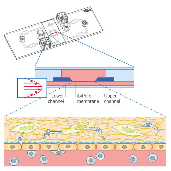
Porous membrane co-culture assays under flow enable the cultivation of two cell layers in separate but interacting compartments. One cell monolayer can be exposed to flow while soluble factors, cell–cell communication, or barrier-related responses are analyzed across the membrane. This assay format is suitable for endothelial and epithelial barrier models, vascular co-culture systems, immune cell interaction studies, transmigration studies, and microscopy of living or fixed cell layers under defined shear stress conditions.
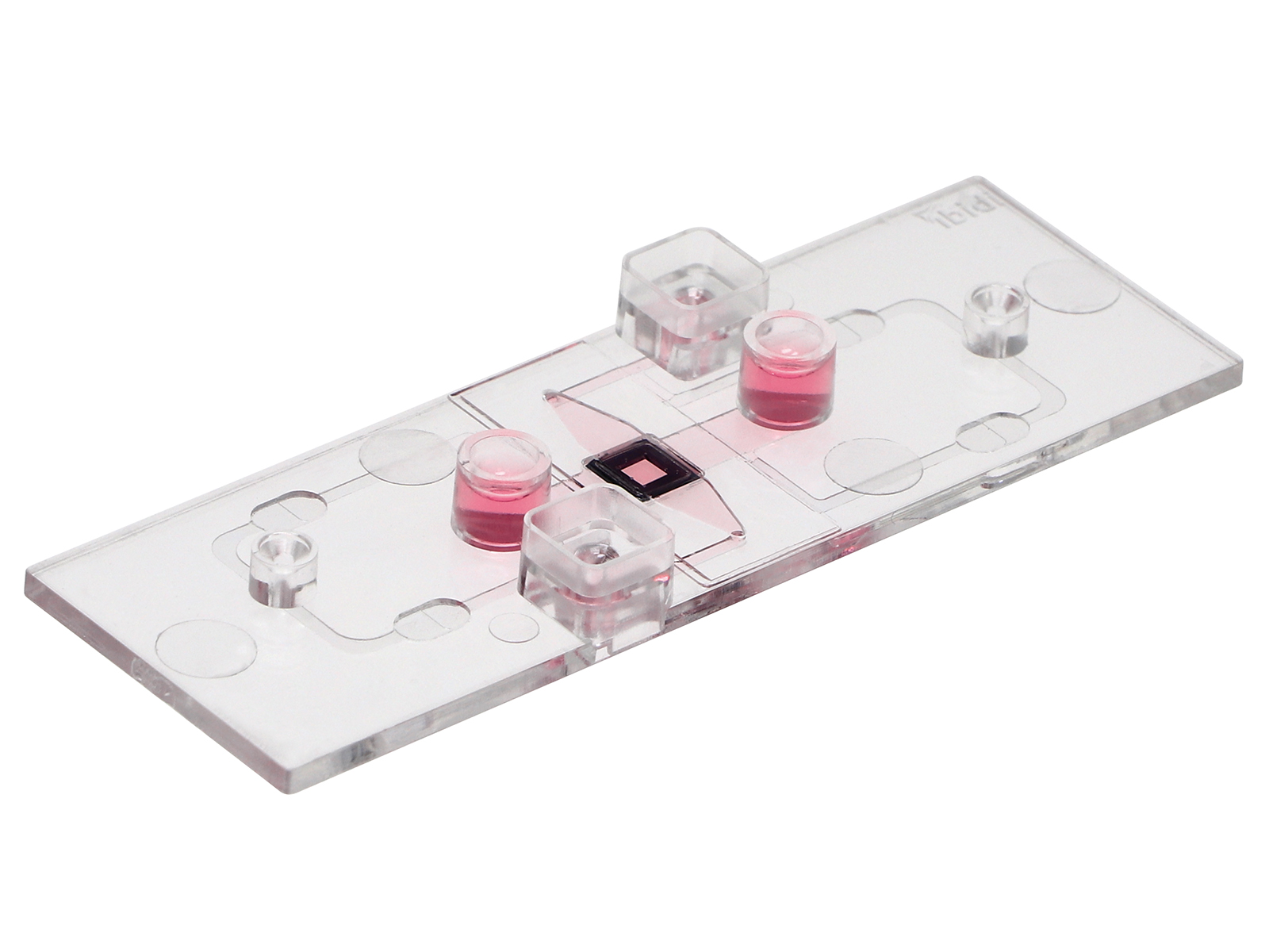
Transmigration of cells across a cell monolayer on the membrane into a 3D gel matrix with embedded cells using the µ-Slide ibiPore SiN.
A Dynamic Blood–Brain Barrier Model for Microscopy and Permeability Analysis
The blood–brain barrier is a highly selective vascular interface that restricts drug entry into the brain, making physiologically relevant in vitro models essential for permeability testing and CNS drug development. In this study by Choublier et al., the authors developed a custom two-compartment BBB device in which hCMEC/D3 brain endothelial cells were cultured on a semi-permeable membrane and exposed to uniform laminar shear stress using the ibidi Pump System. The setup enabled long-term endothelial culture under flow, direct microscopy-based analysis of barrier morphology and junction markers, and permeability measurements under static and dynamic conditions.
Schematic representation of a dynamic blood–brain barrier model for microscopy and permeability analysis. Brain endothelial cells were cultured on a semi-permeable membrane in a custom two-compartment device and perfused under uniform laminar shear stress using the ibidi Pump System. Adapted from: Choublier N, et al. (2021), doi:10.3390/app11125584, licensed under CC BY 4.0.
ibidi Solutions for Barrier Models and Permeability
Selected Publications for Barrier Models and Permeability
A dynamic blood–brain barrier model was established in a custom-made device and perfused using the ibidi Pump System to expose hCMEC/D3 brain endothelial cells to shear stress for microscopy and permeability measurements.
Choublier N, Müller Y, Gomez Baisac L, Laedermann J, de Rham C, Declèves X, Roux A. (2021) Blood–Brain Barrier Dynamic Device with Uniform Shear Stress Distribution for Microscopy and Permeability Measurements. Appl Sci., 10.3390/app11125584.
Read article
HUVEC monolayers were cultured on the µ-Slide ibiPore and exposed to laminar shear stress using the ibidi Pump System to study endothelial barrier integrity, FITC-dextran permeability, and monocyte adhesion/transmigration.
Zhong T, Li Y, He X, Liu Y, Dong Y, Ma H, Zheng Z, Zhang Y. (2020) Adaptation of endothelial cells to shear stress under atheroprone conditions by modulating internalization of vascular endothelial cadherin and vinculin. Ann Transl Med. 8(21):1423, 10.21037/atm-20-3426.
Read article
Neutrophil basement membrane penetration was analyzed using the µ-Slide ibiPore to study Src family kinase-dependent vesicle trafficking during neutrophil extravasation.
Rohwedder I, Kurz ARM, Pruenster M, Immler R, Pick R, Eggersmann T, Klapproth S, Johnson JL, Alsina SM, Lowell CA, Mócsai A, Catz SD, Sperandio M. (2020) Src family kinase-mediated vesicle trafficking is critical for neutrophil basement membrane penetration. Haematologica. 105(7):1845–1856, 10.3324/haematol.2019.225722.
Read article
Neutrophil transmigration was analyzed using the µ-Slide ibiPore to study how Myosin 1f regulates neutrophil migration through 3D environments and basement membrane-like barriers during acute inflammation.
Salvermoser M, Pick R, Weckbach LT, Zehrer A, Löhr P, Drechsler M, Sperandio M, Soehnlein O, Walzog B. (2018) Myosin 1f is specifically required for neutrophil migration in 3D environments during acute inflammation. Blood. 131(17):1887–1898, 10.1182/blood-2017-10-811851.
Read article
Tumor Microenvironment, Extravasation, and Drug Delivery
Perfused tumor microenvironment models can combine endothelial layers, immune cells, tumor spheroids, hydrogel matrices, and dynamic compound delivery. These models are suitable for studying extravasation, immune-tumor interaction, carrier penetration, drug accumulation, and viability under flow.
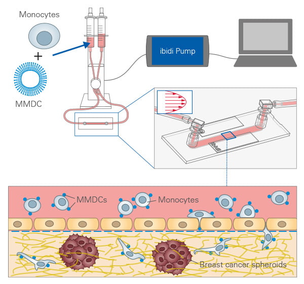
Monocyte-Mediated Drug Delivery in a 3D Tumor Model Under Flow
Immune cell trafficking offers a promising route to improve drug delivery into solid tumors, where vascular barriers and dense extracellular matrices often limit passive nanoparticle accumulation. In this study by Chang et al., the authors developed monocyte-adhesive peptidyl liposomes, referred to as monocyte-mediated drug carriers (MMDCs), designed to hitchhike on circulating monocytes. Using ibidi flow-based assays, they first analyzed the interaction between MMDCs and monocytes under defined shear stress. They then established a 3D tumor microenvironment model with collagen-embedded tumor spheroids and an endothelial barrier to investigate monocyte adhesion, transendothelial migration, and selective MMDC accumulation in cancer-containing matrices under dynamic flow conditions.
Schematic illustration of MMDC-assisted monocyte homing in a 3D tumor microenvironment model under flow. The setup enabled analysis of monocyte adhesion, transendothelial migration, and tumor-directed carrier accumulation. Graphical abstract created by ibidi, based on: Chang CY, et al. (2025), doi:10.1016/j.jconrel.2025.113672.
PD-L1-Targeted Immunoliposomes in a Cervical Cancer-on-a-Chip Model
Cancer-on-a-chip models provide a controlled approach to study how therapeutic nanocarriers behave in a 3D tumor-like environment under perfusion. In this study by Fobian et al., PD-L1-targeted immunoliposomes were investigated in a dynamic cervical cancer model combining tumor spheroids, an extracellular matrix, and defined flow. The setup allowed the authors to assess how flow influences immunoliposome transport, penetration into the 3D matrix, and delivery toward PD-L1-expressing tumor cells in a more physiologically relevant in vitro system.
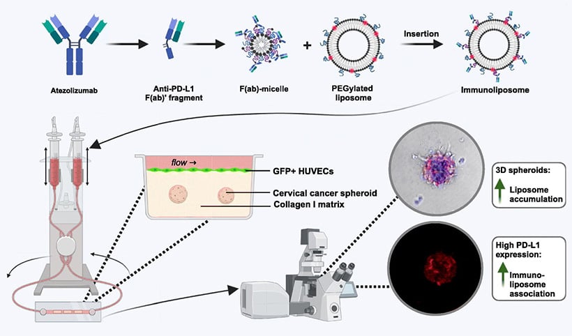
Schematic representation of a dynamic cervical cancer-on-a-chip model for studying PD-L1-targeted immunoliposome delivery. Tumor spheroids were embedded in an extracellular matrix and exposed to controlled perfusion, enabling the analysis of nanocarrier transport, matrix penetration, and tumor-associated accumulation under flow. Reproduced from: Fobian SF, et al. (2025), doi:10.1016/j.jconrel.2025.01.014, licensed under CC BY 4.0.
ibidi Solutions for Tumor Microenvironment, Extravasation, and Drug Delivery
Selected Publications for Tumor Microenvironment, Extravasation, and Drug Delivery
Monocyte-mediated drug carriers were analyzed using the ibidi Pump System, µ-Slide I Luer 3D, µ-Slide I 0.8 Luer, and Collagen Type I to study tumor-targeted drug delivery under physiological flow conditions.
Chang CY, Huang SH, Chen CY, Jian CB, Chang CC, Chang YY, Jung M, Lee HM, Cheng B. (2025) Monocyte-adhesive peptidyl liposomes for harnessing monocyte homing to tumor tissues. J Control Release., 10.1016/j.jconrel.2025.113672.
Read article
PD-L1-targeted immunoliposomes were evaluated in a dynamic cervical cancer-on-a-chip model using the ibidi Pump System and µ-Slide I Luer 3D to study liposome delivery to collagen-embedded tumor spheroids under flow.
Fobian SF, Amin M, Sacchetti A, Seynhaeve ALB, Oei AL, ten Hagen TLM. (2025) Investigating the delivery of PD-L1-targeted immunoliposomes in a dynamic cervical cancer-on-a-chip model. J Control Release. 379:236–250, 10.1016/j.jconrel.2025.01.014.
Read article
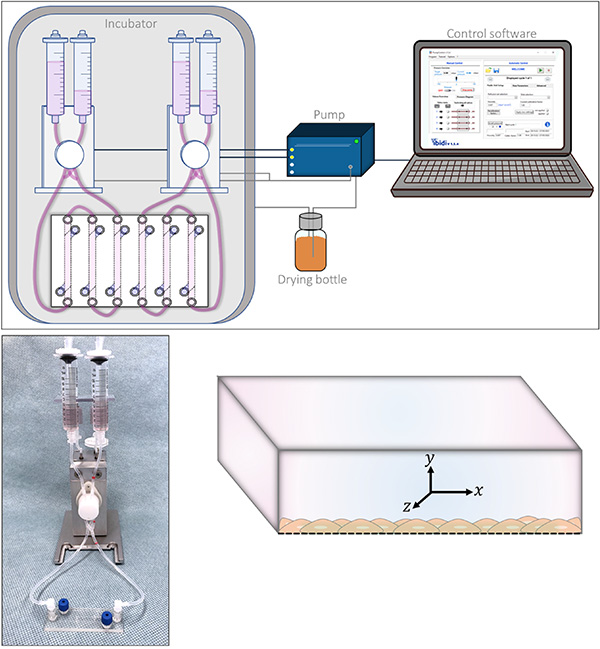
Plan Your ibidi Flow Setup
Wall shear stress assays require a defined flow source, a channel geometry with known dimensions, and stable environmental conditions during imaging or endpoint analysis. For a complete overview of compatible ibidi components and workflow considerations, see the detailed setup explanation.
Frequently Asked Questions About Wall Shear Stress Assay Applications
Which wall shear stress assay format is best for endothelial monolayers?
For standard endothelial monolayers under defined shear stress, a 2D cell monolayer under flow is usually the most direct assay format. It allows controlled flow exposure, microscopy access, long-term flow conditioning, immunofluorescence staining, and downstream endpoint analysis.
When should I use a matrix-based 3D model under flow?
A matrix-based 3D model is useful when extracellular matrix-dependent adhesion, signaling, barrier function, transmigration, or a more tissue-like microenvironment is relevant to the biological question. It can include cells cultured on a matrix, embedded inside a gel, or perfused through the flow channel.
When should I use suspended cells in a flow assay?
Suspended cells in flow are suitable when the experiment should analyze cell rolling, adhesion, cell–cell interaction, immune cell recruitment, or transendothelial migration under defined shear stress. Depending on the model, these cells can interact with a 2D monolayer, an endothelial barrier, or a matrix-based 3D environment.
When should I use a porous membrane co-culture format?
A porous membrane co-culture format is useful when two cell layers should be cultured in separate but interacting compartments. It is suitable for barrier models, endothelial–epithelial interaction studies, immune cell interaction studies, transmigration studies, and microscopy-based analysis under flow.
Which ibidi products are commonly used for wall shear stress assay applications?
Common products include the ibidi Pump System, µ-Slide I Luer Family, µ-Slide VI, µ-Slide y-shaped, µ-Slide I Luer 3D, µ-Slide ibiPore SiN, and compatible matrices such as Collagen Type I. The optimal setup depends on the assay format, flow profile, cell type, matrix requirements, co-culture design, and readout method.