



User Protocol 11:
Protocol for Spheroid Culture, Staining, and Clearing for 3D Imaging
Julian Hofmann1, Louise Ritter2, Selina J. Keppler*1
1 Inflammation and Immunity Lab, TranslaTUM, Institute for Clinical Chemistry and Pathobiochemistry, Klinikum rechts der Isar, Technical University Munich, Germany.
2 ibidi GmbH, Gräfelfing, Germany.
*Corresponding author. E-mail address: selina.keppler@tum.de
This User Protocol describes the steps that are necessary to get accurate 3D images of spheroids after long-term culture under perfusion.
Multicellular spheroids of murine L929 fibroblasts were created in the µ-Slide Spheroid Perfusion. After the formation of spheroids from a cell suspension, perfusion was applied with the ibidi Pump System. Adding the ibidi Pump ensures the optimal nutrition of spheroids during long-term cultivation.
After 1 week in perfusion culture, the spheroids were fixed and stained with phalloidin to visualize the F-actin cytoskeleton. Finally, spheroids were cleared to enhance imaging depth and resolution. We used our EMOVI approach for cost-effective, non-harmful whole-mount imaging, which enables multiplexed antibody-based immunolabeling on fixed spheroids. The clearing of spheroids allows the analysis of the distribution and organization of fibroblasts throughout and even at the center of the spheroid fibroblasts, while at the same time preserving the spheroid’s morphology.
Hofmann J, Gadjalova I, Mishra R, Ruland J and Keppler SJ (2021) Efficient Tissue Clearing and Multi-Organ Volumetric Imaging Enable Quantitative Visualization of Sparse Immune Cell Populations During Inflammation. Front. Immunol. 11, 599495. https://doi.org/10.3389/fimmu.2020.599495.
Hofmann J and Keppler SJ (2021) Tissue clearing and 3D imaging – putting immune cells into context. J Cell Sci 134 (15), jcs258494. https://doi.org/10.1242/jcs.258494.
1. Related Documents
- Instructions µ-Slide Spheroid Perfusion
- Instructions ibidi Pump System
- Application Note 31: Serial Connection of µ-Slide VI 0.4
2. Materials and Reagents
Cells and Reagents
- L929 Fibroblasts (DSMZ, no. ACC 2)
- Accutase (A1110501, Gibco)
- Cell Culture Medium RPMI-1640 (Gibco, 21875034)
- Fetal Calf Serum (FCS, Gibco, 10270106)
- Intracellular (IC) Fixation Buffer (eBioscience, 00-8222-49)
- Dulbecco’s Phosphate Buffer Saline (PBS, Sigma, D8537)
- Normal Mouse Serum (Jackson, 015-000-120)
- Normal Donkey Serum (Jackson, AB_2337258)
- Triton X-100 (Alfa Aesar, A16046)
- Flash Phalloidin™ Green 488 (Phalloidin; stock: 500x, Thermo Fisher Scientific, 46410)
- 4',6-Diamidino-2-Phenylindole (DAPI, stock conc. 20 µg/mL, Sigma-Aldrich, D9542)
- Deionized water (diH2O)
- Isopropanol (Carl Roth, CP41.2)
- Ethyl Cinnamate (ECi) (Sigma, W243000)
3. Buffers and Solutions
Culture Medium
- Prepare freshly and store at 4°C for up to one week
- Basal Medium RPMI-1640 (Gibco, 21875034)
- 10% FCS (Gibco, 10270106)
Blocking and Staining Buffer
- PBS
- 1% (v/v) FCS
- 1% (v/v) Normal Mouse Serum
- 1% (v/v) Normal Goat Serum
- 0.3% (v/v) Triton X-100
Staining Solution
- Blocking and Staining Buffer
- 1:500 Phalloidin (final conc. 1x)
- 1:10 DAPI (final conc. 2 µg/mL)
30% / 50% / 70% (v/v) Isopropanol Dilutions
- Mix appropriate volumes of diH2O and isopropanol
- Pre-chill to 4°C before use
4. Equipment
- ibidi Pump System (ibidi, 10902)
- ibidi µ-Slide Spheroid Perfusion with Bioinert surface passivation (ibidi, 80350)
- ibidi Serial Connector for µ-Slides (ibidi, 10830)
- ibidi Perfusion Set Blue (ibidi, 10961)
- Laminar Flow Hood
- Incubator, 37°C and 5% CO2
- Neubauer hemocytometer
- Pipettes
- Inverted confocal microscope (here: Leica TCS SP8)
- Leica Application Suite X
- LIGHTNING (deconvolution software)
- Imaris v9.5
5. Procedure
Part I: Spheroid Formation and Culture in the µ-Slide Spheroid Perfusion
Please read the Instructions before working with the µ-Slide Spheroid Perfusion. Perform all steps under sterile conditions.
Before starting the experiment, prepare the L929 fibroblasts in a standard cell culture flask (e.g., T75) with the cells adherent at the bottom. The cells should be healthy and optimally subconfluent on the day of the experiment.
Important Note: Equilibrate all required materials, such as µ-Slides, culture medium, and tubing (Perfusion Sets), overnight inside the incubator at 37°C and 5% CO2. Equilibration is essential for keeping air bubbles from emerging over time.
- Inject 60 µl cell-free culture medium into each channel of the µ-Slide Spheroid Perfusion (closed with the supplied coverslip according to the Instructions)
- Incubate for 2 h at 37°C in the incubator. Always close the lid during incubation.
- Treat the cultured L929 cells with Accutase for 1–2 minutes for detachment.
- Harvest the cell suspension, centrifuge, and dilute it in culture medium; the amount depends on the required cell concentration.
- Count the cells and adjust to a final concentration of 5 x 10⁵ cells/ml.
- Flush the channels of the µ-Slide Spheroid Perfusion with cell-free culture medium to remove potential air bubbles.
- Pipet 45 µl cell suspension directly into each channel.
- Incubate for 1 h at 37°C for the cells to settle in the niche.
- Fill the reservoirs of the µ-Slide Spheroid Perfusion with 60 µl cell-free culture medium.
- Incubate overnight at 37°C for spheroid formation.
- Check the channels for air bubbles. If any bubbles are present in the channel, carefully flush the channel with cell-free culture medium.
- Connect the 3 channels of the µ-Slide Spheroid Perfusion using ibidi Serial Connectors (see Application Note 31: Serial Connection of the Six Channels of the µ-Slide VI 0.4).
- Prepare the ibidi Pump System, Fluidic Unit, and Perfusion Set according to the Instructions.
- Connect the µ-Slide Spheroid Perfusion to the ibidi Pump System and start the flow. Use the lowest possible flow rate (5 mbar air pressure resulting in a flow rate of 0.74 ml/min). The experiment is conducted until the spheroids have the desired maturation state, in this case, after 7 days.
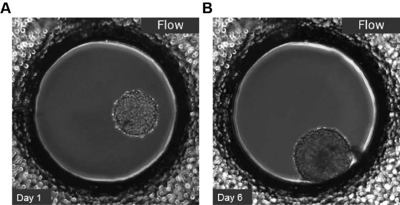
Figure 1. L929 fibroblasts show spheroid formation in the µ-Slide Spheroid Perfusion. Examples of day 1 (A) and day 6 (B), perfusion with the ibidi Pump System, 0.74 ml/min. Phase contrast microscopy, 10x objective lens, well diameter 800 µm.
Part II: Staining and Clearing of L929 Spheroids
Staining is performed directly in the µ-Slide Spheroid Perfusion. Before starting the staining, please read the Instructions and follow the recommendations for general pipetting and changing solutions in the slide.
Important Note: ECi can be harmful to plastic materials. We cannot generally guarantee that ECi is compatible with the ibidi labware. Please use the ibidi Free Sample Program to test ECi solution compatibility with plastic labware.
- When L929 spheroid cultures have reached the desired maturation state (here, after 7 days), carefully disconnect the µ-Slide Spheroid Perfusion from the ibidi Pump System.
- Fix the spheroids with cold IC Fixation Buffer for 10 min at RT.
- Incubate spheroids in Blocking and Staining Buffer for 1 h at RT.
- Incubate spheroids with the Staining Solution overnight at 37°C. From this point forward, keep the slide protected from light. Figure 2A shows spheroid imaging before clearing.
- On the next day, wash the cells once with Blocking and Staining Buffer.
- Sequentially dehydrate the spheroids by incubating in ascending 30%, 50%, and 70% isopropanol dilutions for 15 min each on ice.
- Incubate spheroids twice with pure isopropanol for 15 min on ice.
- Remove slide from ice and let it warm up to RT.
- Perform the clearing: perfuse twice with pure ECi at RT. At this stage, spheroids can be stored protected from light for several weeks without significant loss of fluorescence.
- Image spheroids using a confocal microscope (see Figure 2).
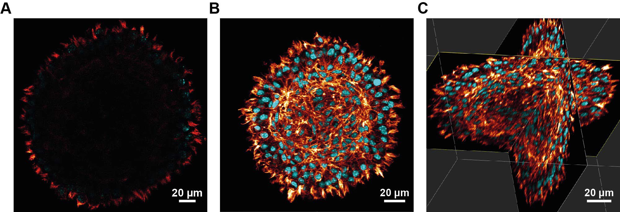
Figure 2. Confocal microscopy of a stained L929 spheroid before and after clearing (red: phalloidin, cyan: DAPI).
(A) Spheroid before clearing (after step 4 in this protocol). Note that due to light scattering and absorption only peripheral cells are visible, whereas the cells in the center cannot be seen. (B, C) Spheroid after clearing. Note that the size difference between pre- and post-clearing stems from the dehydration and clearing process, cross-section taken at the same position. (B) Cross-section (C) Volumetric/3D view. Clearing of the spheroids allows the visualization of cells even at the center of the spheroid fibroblasts while at the same time preserving the spheroid’s morphology. Counting of cell nuclei would even allow the determination of cell numbers forming this spheroid.
Movie: View the direct comparison of a stained spheroid before and after clearing as a movie here (z-stack of 440 slices at a slice spacing of 0.36 µm).
This User Protocol is an ibidi peer-reviewed protocol from an actual user. ibidi does not guarantee its functionality or reproducibility. For this User Protocol, ibidi provides only limited support. Please contact the corresponding author for detailed information.
For research use only.