



User Protocol 07:
Protocol for DRG Neuron Preparation
Flavia Millesi*, Tamara Weiss, Christine Radtke
*Corresponding author: flavia.millesi@meduniwien.ac.at
Nerve cells account for one of the most demanding and complex cell types in culture. Nevertheless, they are indispensable for neuroscience research. Among these nerve cells are dorsal root ganglia (DRG) neurons. They represent sensory neurons of the peripheral nervous system. The successful culture of DRG neurons is especially important to study pain reception and, as in our case, peripheral nerve regeneration.
In this protocol, we show how to harvest, culture, and identify rat DRG neurons. From the DRGs of adult Sprague-Dawley rats, neurons were isolated using a Percoll-gradient. Afterwards, cells were seeded on the µ-Slide 8 Well and cultured for several days. Upon fixation, multicolor immunofluorescence staining panels helped to identify DRG neurons amongst other cells like Schwann cells and fibroblasts.
1. Materials and Reagents
Reagents:
- 1x Dulbecco’s Phosphate Buffer Saline (PBS)
- 20% Percoll solution
- MEMα medium
- Neurobasal medium
- Laminin
- Poly-D-lysine
- 1% penicillin/streptomycin (Pen/Strep)
- Collagenase type IV
- Dispase II
- Calcium chloride
- 2.5% 4-(2-hydroxyethyl)-1-piperazineethanesulfonic acid buffer solution (HEPES)
- Fetal calf serum (FCS)
- recombinant NGF
- 4.5% formaldehyde solution
- 1% bovine serum albumin (BSA)
- Goat serum
- Triton X-100
- 4',6-Diamidino-2-phenylindole (DAPI)
- FluoromountG mounting medium
- Anti-S100
- Anti-β-3-tubulin (TUJ1)
- Anti-Vimentin (VIME)
2. Solutions and Culture Media
Digestion solution:
Note: prepare fresh and use immediately
- MEMα medium
- Pen/Strep (f.c. 1%)
- FCS (f.c. 10%)
- Sodium Pyruvate Solution (f.c. 1%)
- HEPES (f.c. 2.5%)
- Collagenase type IV (f.c. 0.125% (w/v))
- Dispase II (f.c. 1.25 U/ml)
- Calcium chloride (f.c. 3 mM)
DRG culture medium:
Note: prepare fresh, keep at 4°C, and use no longer than one week
- NeurobasalTM-A medium
- 10 ng/ml recombinant NGF
- 1x B27 supplement (f.c. 1x)
- L-glutamine (f.c. 2 mM)
Coating solutions:
Note: PDL and laminin solutions can be stored at 4°C for 4 weeks
PDL solution:
Prepare PDL solution (f.c. 0.01%) in ddH2O
Laminin solution:
Prepare laminin solution (f.c. 4.8 μg/ml) diluted in 1x PBS
Blocking and permeabilization (Block/Perm) solution:
- 1x PBS
- BSA (f.c. 1%)
- goat serum (f.c. 5%)
- Triton X-100 (f.c. 0.3%)
Antibody staining solution:
- 1x PBS
- BSA (f.c. 1%)
- goat serum (f.c. 1%)
- Triton X-100 (f.c. 0.1%)
- Primary and secondary antibodies (see Table 1)
DAPI working solution:
- 1x PBS
- DAPI (f.c. 50 µg/ml)
Table 1. List of used primary and secondary antibodies
| Primary Antibodies | |||
|---|---|---|---|
| Antigen | Species | Dilution | Company |
| S100 | Rabbit | 1:200 | DAKO |
| Vimentin (VIME) | Chicken | 1:300 | Thermo Fisher |
| β-3-tubulin (TUJ1) | Mouse | 1:100 | Santa Cruz |
| Secondary Antibodies | |||
| Fluorophore | Target species | Dilution | Company |
| AF594 | Rabbit | 1:400 | Thermo Fisher |
| DL650 | Chicken | 1:400 | Thermo Fisher |
| AF488 | Mouse | 1:400 | Thermo Fisher |
3. Equipment
- Laminar Flow Hood
- Incubator, 37°C and 5% CO2
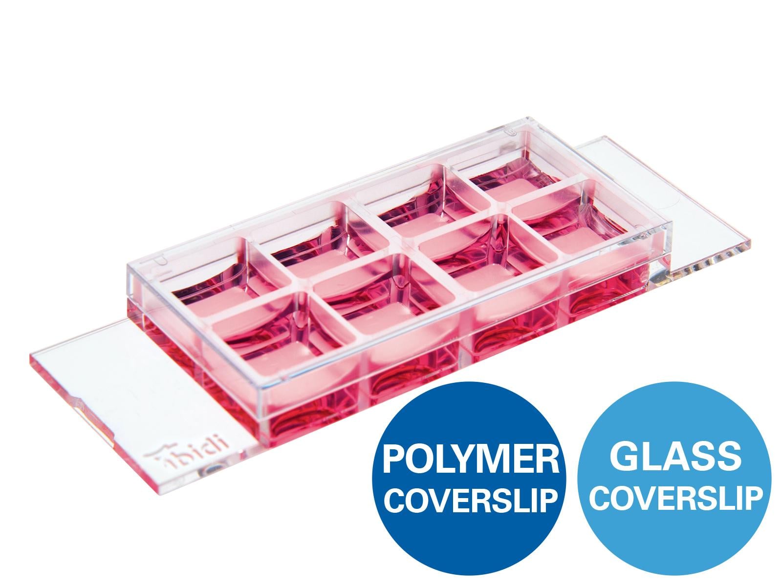
- µ-Slide 8 Well, ibiTreat (ibidi, 80826)
- Pipettes
- Centrifuge
- 15 ml conical bottom centrifuge tubes
- Inverted phase-contrast microscope (here: Nikon Eclipse Ts2R) + Software
- Inverted confocal microscope (here: Leica SPX8) + Software
4. Animals
Adult Sprague-Dawley rats were euthanized in compliance with the Austrian Animal Testing Law (TVG 2012, §2, 1.c) and Article 3 of the Directive 2010/63/EU of The European Parliament and of the Council on the Protection of Animals Used for Scientific Purposes.
5. Procedure
The herein described procedure describes the treatment of DRGs to culture and characterize primary sensory neurons for experimentation. The protocol refers to a harvest of lumbar DRGs derived from the spine of one adult Sprague-Dawley rat. The achieved number of cells is usually sufficient to fill the eight chambers of one µ-Slide 8 Well.
Part I: Establishment of DRG Cultures
- Harvest 6 lumbar DRGs from a euthanized, adult Sprague-Dawley rat and place in 1x PBS supplemented with 1% Pen/Strep on ice (see Figure 1a).
- Under a laminar flow hood, transfer DRGs into the digestion solution and place in an incubator overnight.
- For coating, add 100 µl PDL solution per well of ibidi µ-Slides and incubate at room temperature for 2 hours. Afterwards, remove the PDL solution and add 100 µl laminin solution per well and leave slides overnight in the incubator.
- On the next day, carefully resuspend until the tissue has dissolved and stop the digest by adding MEMα medium and centrifuge cell suspension at 200 x g for 5 minutes.
- Mix 1 ml Percoll solution with 5 ml MEMα medium to generate a 20% Percoll solution.
- Resuspend cells in 1 ml NeurobasalTM-A medium. Pipette the cell solution very slowly on top of the 6 ml 20% Percoll solution and centrifuge at 450 x g for 8 minutes.
- To wash the DRGs, resuspend cells in 1 ml NeurobasalTM-A medium and centrifuge at 1230 x g for 2 minutes.
- In the meanwhile, prepare ibiTreat µ-Slide for cell seeding: Remove laminin and add 150 µl DRG Medium per well.
- Resuspend the cell pellet in 1 ml DRG culture medium and seed 125 µl of DRG cell suspension per coated well of ibiTreat µ-Slide chambered coverslips.
- Outgrowth of neuritic processes from DRG cell bodies can be observed from day 1 and will increase with culture time (Figure 1b-d). Note that DRG cultures also contain Schwann cells and fibroblast-like cells (Figure 1b-d). Medium should be changed three times a week.
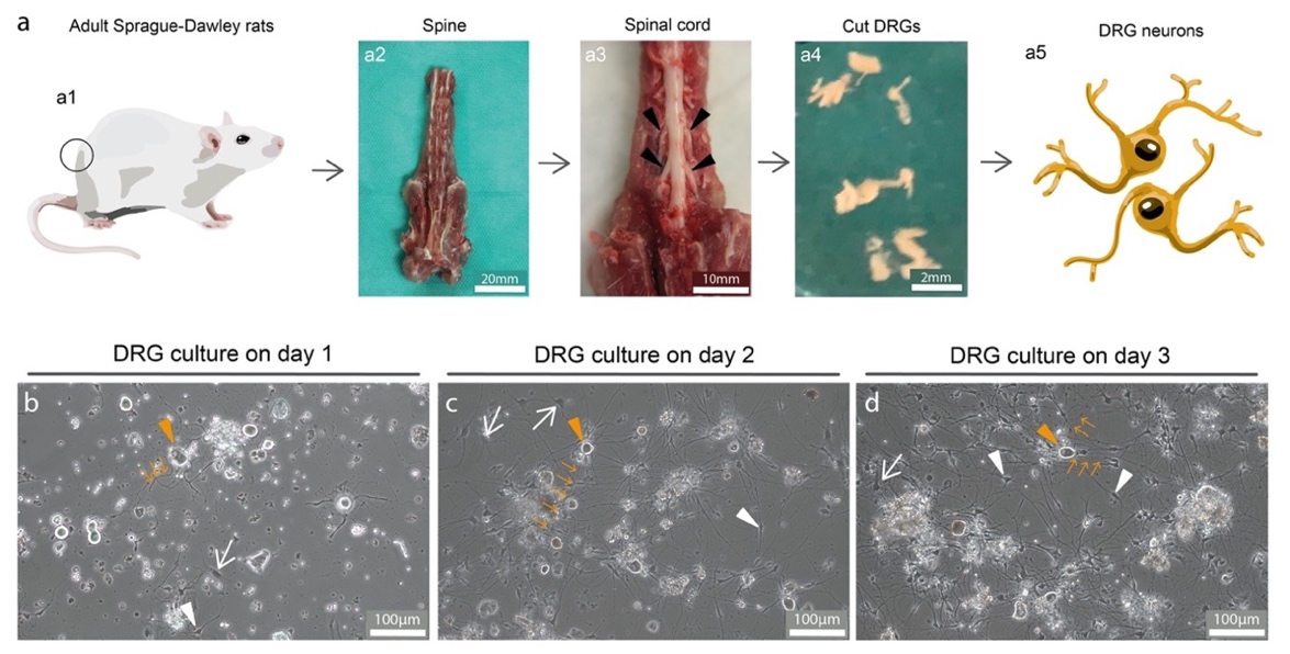
Figure 1. Establishment of primary DRG neuron culture. a) Schema of rat DRG isolation. From adult Sprague-Dawley rats (a1), the spine was removed (a2) and the spinal cord with DRGs was exposed (arrowhead in a3). Isolated DRGs (a4) were digested overnight and seeded on the next day. Representative phase contrast images of a DRG culture on day 1 (b), day 2 (c) and day 3 (d) showing DRG neurons (orange arrowheads), Schwann cells (white arrowheads) and fibroblast-like cells (white arrows). Neurite outgrowth was visible from day 1 (orange arrows).
Part II: Immunostainings of DRG Cultures
Please find antibody details in Table 1. Each washing step involves three washes with 1x PBS for 5 min. The staining procedure is conducted at room temperature unless noted otherwise.
- When DRG cultures have reached the desired maturation state, wash cells once with 1x PBS. Fix cells by adding 4.5% formaldehyde solution for 20 minutes and wash with 1x PBS. Note: the fixation step should be performed under a chemical fume hood. Fixed cells can be stored in 1x PBS at 4°C for about two weeks before the immunostainings are performed.
- For immunostaining of DRG cultures, block and simultaneously permeabilize cells by applying 100 µl of block/perm solution for 10 minutes.
- Incubate cells with 100 µl antibody staining solution containing primary antibodies overnight at 4°C.
- On the next day, wash cells and add 100 µl secondary antibodies dissolved in the antibody staining solution for 1 hour.
- Wash cells and incubate with DAPI working solution for 10 minutes.
- After another washing step, add 250 µl FluoromountG mounting medium per well and store slides at 4°C.
- Image cells using a confocal microscope and identify TUJ1 positive sensory neurons, S100 and VIME positive Schwann cells and VIME positive fibroblast-like cells within the DRG cultures (see Figure 2).
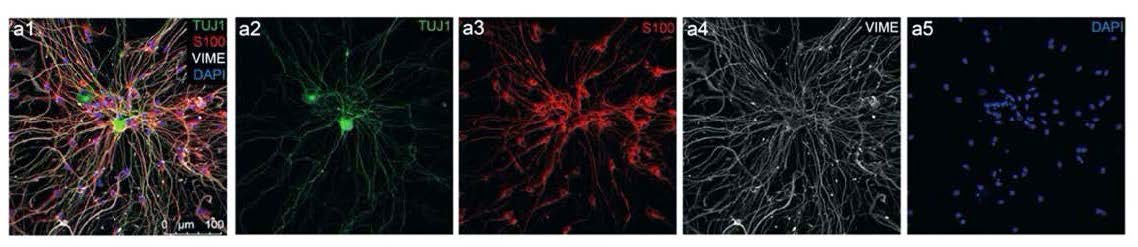
Figure 2. Confocal image analysis of immunostained DRG cultures. DRG cultures were stained for neuronal marker TUJ1 (a2), Schwann cell marker S100 (a3), intermediate filament vimentin (VIME, a4) and DAPI (a5), merged channels are shown in a1. DRG neurons showed positive staining for the TUJ1 in the cell body and along the neurites, but were negative for S100, whereas Schwann cells were positive for S100 and VIME, but negative for TUJ1. Fibroblast-like cells were positive for VIME, but negative for TUJ1 and S100.
6. References
Weiss T, Taschner-Mandl S, Ambros PF, Ambros IM. Detailed Protocols for the Isolation, Culture, Enrichment and Immunostaining of Primary Human Schwann Cells. Methods Mol Biol. 2018;1739:67-86. doi: 10.1007/978-1-4939-7649-2_5. PMID: 29546701. (Methods are based on and adapted from this protocol)
Millesi F, Weiss T, Mann A, Haertinger M, Semmler L, Supper P, Pils D, Naghilou A, Radtke C. Defining the regenerative effects of native spider silk fibers on primary Schwann cells, sensory neurons, and nerve-associated fibroblasts. FASEB J. 2021 Feb;35(2):e21196. doi: 10.1096/fj.202001447R. Epub 2020 Nov 19. PMID: 33210360; PMCID: PMC7894153. (Methods described in this protocol were published in this article)
This User Protocol is an ibidi peer-reviewed protocol from an actual user. ibidi does not guarantee its functionality or reproducibility. For this User Protocol, ibidi provides only limited support. Please contact the corresponding author for detailed information.
For research use only.