



User Protocol 13:
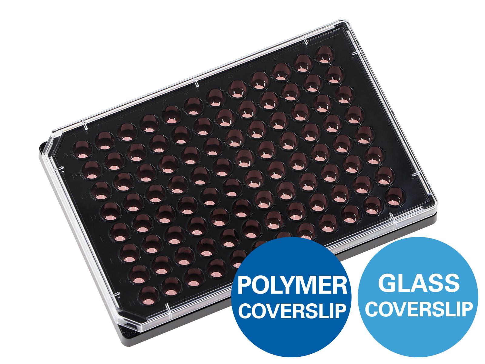
Protocol for Immunofluorescent Staining of iPS Cell-Derived Dopaminergic Neurons in the ibidi µ-Plate 96 Well Square
Asuka Morizane*
Kobe City Medical Center General Hospital, Kobe, Japan.
*Corresponding author. E-mail address: asuka_morizane@kcho.jp
Induced pluripotent stem (iPS) cells are commonly used in neuroscience research. Derived from human somatic cells after converting them into proliferative pluripotent cells, they can be further differentiated into neurons with a midbrain dopaminergic phenotype. These cells play a significant role in understanding the cellular mechanisms underlying neurological diseases and show a high potential in treating neurodegenerative disorders like Parkinson’s disease.
This User Protocol describes the process of immunofluorescent staining of iPS cell-derived dopaminergic neurons in the ibidi µ-Plate 96 Well Square. Please refer to other protocols for the induction of dopaminergic neurons from iPS cells.
1. Related Documents
2. Materials and Reagents
Cells and Reagents
- iPS cell-derived dopaminergic neurons at differentiation day 30 (original iPS cell line: 1231A3)
- iMatrix-511 (Nippi)
- Neurobasal Medium (Thermo Fisher Scientific, 21103049)
- B27 (Thermo Fisher Scientific, 12587010)
- Glutamax-I (Thermo Fisher Scientific, A1286001)
- Glial cell-derived neurotrophic factor (GDNF, Wako)
- Ascorbic acid (Wako)
- Brain-derived neurotrophic factor (BDNF, Wako)
- dibutyryl cAMP (dbcAMP, Sigma-Aldrich)
- Y-27632 (Fujifilm)
- Paraformaldehyde (PFA)
- Phosphate-buffered Saline (PBS)
- Normal Donkey Serum (NDS)
- Triton X-100
- Tween 20
Buffers and Solutions
Neural Differentiation Medium
- Neurobasal Medium
- B27 (10 ml for 500 ml of Neurobasal Medium)
- 2 mM Glutamax-I
- 10 ng/ml GDNF
- 200 µM ascorbic acid
- 20 ng/ml BDNF
- 400 µM dbcAMP
- 20 µM Y-27632 (added to the medium for seeding the cells to inhibit apoptosis induced by mechanical stress)
Blocking Buffer
- PBS
- 2–2.5% NDS
- 0.3% (v/v) Triton X-100
Washing Buffer
- PBS
- 0.05% (v/v) Tween 20
Equipment
- µ-Plate 96 Well Square, ibiTreat (ibidi, 89626)
- Laminar Flow Hood
- Incubator, 37°C and 5% CO2
- Pipettes and respective tips
- Inverted fluorescent microscope (here: BZ-X710 microscope, Keyence)
| Antibody/stain | Manufacturer | Cat. No. | Working concentration |
|---|---|---|---|
| Primary antibody | |||
| Rabbit anti-Tyrosine hydroxylase (TH) antibody | Millipore | AB152 | 1:500 |
| Mouse anti-TubβIII antibody | BioLegend | 801201 | 1:1000 |
| Goat anti-FoxA2 antibody | R&D | AF2400 | 1:500 |
| Secondary antibody | |||
| Alexa Fluor series donkey Donkey anti-Mouse Alexa Fluor 488 Donkey anti-Rabbit Alexa Fluor 594 Donkey anti-Goat Alexa Fluor 594 Donkey anti-Mouse Alexa Fluor 647 | Invitrogen | A21202 A21207 A11058 A31571 | 1:400 |
| Counterstaining | |||
| DAPI | Nacalai Tesque | 19178-91 | 1:48,000 |
3. Procedure
Please read the Instructions before working with the µ-Plate 96 Well Square.
3.1 Sample Preparation
- Perform all steps under sterile conditions.
- Coat the plate with 0.55 µl (0.28 µg) iMatrix-511 in 90 µl PBS per well for 1–24 hours at 37°C.
- Plate iPS-derived dopaminergic progenitor cells onto the µ-Plate 96 Well Square at a density of 1.7 x 10⁵ cells in a volume of 180 µl Neural Differentiation Medium per well.
- Incubate iPS cells at 37°C for seven days (i.e., 37 days of differentiation in total).
- Gently exchange half of the medium every two to three days. We do not recommend a total-volume medium exchange since neural cells easily detach from the surface.
For the following steps, 90 µl of the respective buffers are used per well. If the cells are kept in the refrigerator for an extended period, it is recommended to add 180 µl PBS per well to accommodate for evaporation.
3.2 Fixation
- Aspirate the cell culture medium.
- Fill with 4% PFA and incubate the cells for 30 minutes at 4°C.
- Aspirate PFA.
- Fill the wells with PBS.
It is recommended to immediately continue with the next step. However, fixed cells can be stored for a short time at 4°C.
3.3 Permeabilization and Blocking
- Aspirate PBS.
- Incubate the cells with Blocking Buffer for one hour at room temperature.
3.4 Primary Antibody Staining
- Prepare the primary staining solution by diluting the primary antibody in Blocking Buffer.
- Discard the Blocking Buffer and immediately move to the next step. Do not let the cells dry out.
- Add the primary staining solution to the cells and incubate overnight at room temperature.
- Rinse the cells three times with Washing Buffer.
3.5 Secondary Antibody Staining
- Prepare the secondary staining solution by diluting the secondary antibodies in Blocking Buffer.
- Aspirate the Washing Buffer.
- Add the secondary staining solution to the cells and incubate for 1 hour at room temperature.
- Rinse the cells two times with Washing Buffer.
3.6 Counterstaining
- Prepare the counterstaining solution by diluting DAPI in PBS (1:48,000).
- Aspirate the Washing Buffer.
- Add the counterstaining solution to the cells and incubate for 10 minutes at room temperature.
- Rinse the cells once with Washing Buffer.
- Fill the wells with PBS and keep the samples in the dark until image acquisition.
4. Image Acquisition
Image the cells using a fluorescence microscope according to the manufacturer’s protocol.
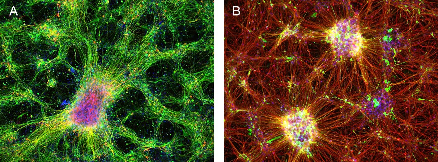
Figure 1. Immunofluorescence image of dopaminergic neurons derived from human induced pluripotent stem cells (iPSCs) in an ibidi µ-Plate 96 Well Square. (A) The image shows the neurite extension with the expression of β-III Tubulin (green) and tyrosine hydroxylase (red). DAPI (blue) was used for nuclear staining. (B) The image shows the neurite extension with the expression of tyrosine hydroxylase (green), β-III Tubulin (red), and Foxa2 (blue). Images were acquired using a BZ-X710 microscope (Keyence).
This User Protocol is an ibidi peer-reviewed protocol from an actual user. ibidi does not guarantee its functionality or reproducibility. For this User Protocol, ibidi provides only limited support. Please contact the corresponding author for detailed information.
For research use only.